Magnesium »
PDB 4y8n-4yes »
4ydh »
Magnesium in PDB 4ydh: The Structure of Human FMNL1 N-Terminal Domains Bound to CDC42
Protein crystallography data
The structure of The Structure of Human FMNL1 N-Terminal Domains Bound to CDC42, PDB code: 4ydh
was solved by
S.Kuhn,
K.Anand,
M.Geyer,
with X-Ray Crystallography technique. A brief refinement statistics is given in the table below:
| Resolution Low / High (Å) | 47.70 / 3.80 |
| Space group | P 21 21 21 |
| Cell size a, b, c (Å), α, β, γ (°) | 87.600, 102.100, 170.600, 90.00, 90.00, 90.00 |
| R / Rfree (%) | 20.8 / 28.1 |
Magnesium Binding Sites:
The binding sites of Magnesium atom in the The Structure of Human FMNL1 N-Terminal Domains Bound to CDC42
(pdb code 4ydh). This binding sites where shown within
5.0 Angstroms radius around Magnesium atom.
In total 2 binding sites of Magnesium where determined in the The Structure of Human FMNL1 N-Terminal Domains Bound to CDC42, PDB code: 4ydh:
Jump to Magnesium binding site number: 1; 2;
In total 2 binding sites of Magnesium where determined in the The Structure of Human FMNL1 N-Terminal Domains Bound to CDC42, PDB code: 4ydh:
Jump to Magnesium binding site number: 1; 2;
Magnesium binding site 1 out of 2 in 4ydh
Go back to
Magnesium binding site 1 out
of 2 in the The Structure of Human FMNL1 N-Terminal Domains Bound to CDC42
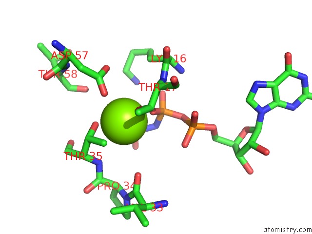
Mono view
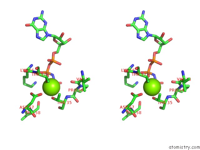
Stereo pair view
Mono view
Stereo pair view
A full contact list of Magnesium with other atoms in the Mg binding
site number 1 of The Structure of Human FMNL1 N-Terminal Domains Bound to CDC42 within 5.0Å range:
|
Magnesium binding site 2 out of 2 in 4ydh
Go back to
Magnesium binding site 2 out
of 2 in the The Structure of Human FMNL1 N-Terminal Domains Bound to CDC42
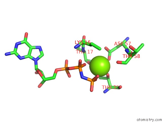
Mono view
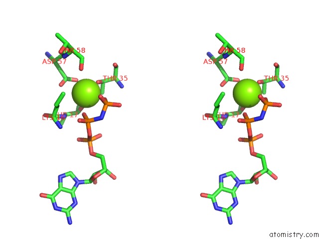
Stereo pair view
Mono view
Stereo pair view
A full contact list of Magnesium with other atoms in the Mg binding
site number 2 of The Structure of Human FMNL1 N-Terminal Domains Bound to CDC42 within 5.0Å range:
|
Reference:
S.Kuhn,
C.Erdmann,
F.Kage,
J.Block,
L.Schwenkmezger,
A.Steffen,
K.Rottner,
M.Geyer.
The Structure of FMNL2-CDC42 Yields Insights Into the Mechanism of Lamellipodia and Filopodia Formation. Nat Commun V. 6 7088 2015.
ISSN: ESSN 2041-1723
PubMed: 25963737
DOI: 10.1038/NCOMMS8088
Page generated: Sat Sep 28 23:14:19 2024
ISSN: ESSN 2041-1723
PubMed: 25963737
DOI: 10.1038/NCOMMS8088
Last articles
Zn in 9J0NZn in 9J0O
Zn in 9J0P
Zn in 9FJX
Zn in 9EKB
Zn in 9C0F
Zn in 9CAH
Zn in 9CH0
Zn in 9CH3
Zn in 9CH1