Magnesium »
PDB 4yf4-4you »
4ygi »
Magnesium in PDB 4ygi: Crystal Structure of SUVH5 Sra Bound to Fully Hydroxymethylated Cg Dna
Enzymatic activity of Crystal Structure of SUVH5 Sra Bound to Fully Hydroxymethylated Cg Dna
All present enzymatic activity of Crystal Structure of SUVH5 Sra Bound to Fully Hydroxymethylated Cg Dna:
2.1.1.43;
2.1.1.43;
Protein crystallography data
The structure of Crystal Structure of SUVH5 Sra Bound to Fully Hydroxymethylated Cg Dna, PDB code: 4ygi
was solved by
E.Rajakumara,
with X-Ray Crystallography technique. A brief refinement statistics is given in the table below:
| Resolution Low / High (Å) | 33.96 / 2.60 |
| Space group | P 42 21 2 |
| Cell size a, b, c (Å), α, β, γ (°) | 76.980, 76.980, 72.111, 90.00, 90.00, 90.00 |
| R / Rfree (%) | 23.5 / 28.3 |
Magnesium Binding Sites:
The binding sites of Magnesium atom in the Crystal Structure of SUVH5 Sra Bound to Fully Hydroxymethylated Cg Dna
(pdb code 4ygi). This binding sites where shown within
5.0 Angstroms radius around Magnesium atom.
In total 3 binding sites of Magnesium where determined in the Crystal Structure of SUVH5 Sra Bound to Fully Hydroxymethylated Cg Dna, PDB code: 4ygi:
Jump to Magnesium binding site number: 1; 2; 3;
In total 3 binding sites of Magnesium where determined in the Crystal Structure of SUVH5 Sra Bound to Fully Hydroxymethylated Cg Dna, PDB code: 4ygi:
Jump to Magnesium binding site number: 1; 2; 3;
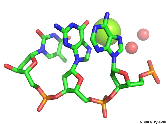
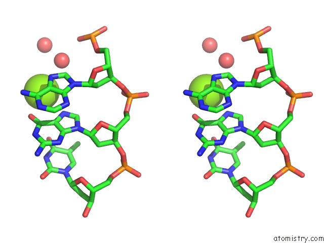
Magnesium binding site 1 out of 3 in 4ygi
Go back to
Magnesium binding site 1 out
of 3 in the Crystal Structure of SUVH5 Sra Bound to Fully Hydroxymethylated Cg Dna
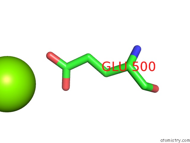
Mono view
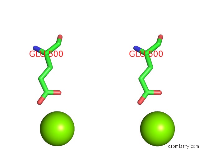
Stereo pair view
Mono view
Stereo pair view
A full contact list of Magnesium with other atoms in the Mg binding
site number 1 of Crystal Structure of SUVH5 Sra Bound to Fully Hydroxymethylated Cg Dna within 5.0Å range:
|
Magnesium binding site 2 out of 3 in 4ygi
Go back to
Magnesium binding site 2 out
of 3 in the Crystal Structure of SUVH5 Sra Bound to Fully Hydroxymethylated Cg Dna
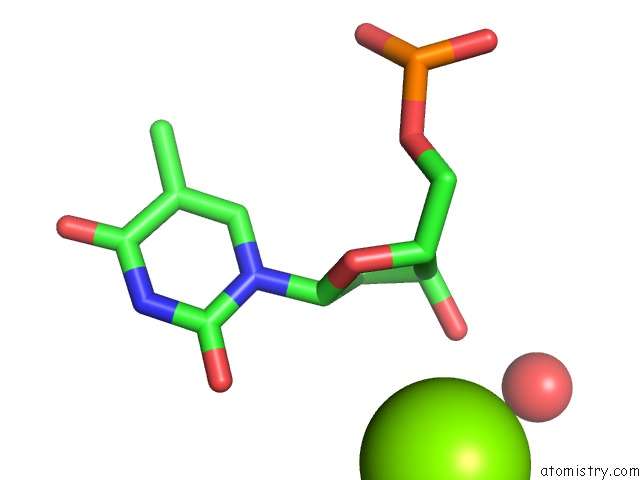
Mono view
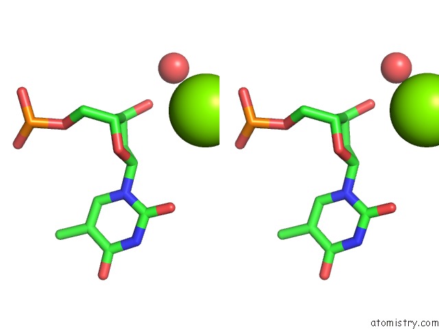
Stereo pair view
Mono view
Stereo pair view
A full contact list of Magnesium with other atoms in the Mg binding
site number 2 of Crystal Structure of SUVH5 Sra Bound to Fully Hydroxymethylated Cg Dna within 5.0Å range:
|
Magnesium binding site 3 out of 3 in 4ygi
Go back to
Magnesium binding site 3 out
of 3 in the Crystal Structure of SUVH5 Sra Bound to Fully Hydroxymethylated Cg Dna
Mono view
Stereo pair view
Mono view
Stereo pair view
A full contact list of Magnesium with other atoms in the Mg binding
site number 3 of Crystal Structure of SUVH5 Sra Bound to Fully Hydroxymethylated Cg Dna within 5.0Å range:
|
Reference:
E.Rajakumara,
N.K.Nakarakanti,
M.A.Nivya,
M.Satish.
Mechanistic Insights Into the Recognition of 5-Methylcytosine Oxidation Derivatives By the SUVH5 Sra Domain Sci Rep V. 6 20161 2016.
ISSN: ESSN 2045-2322
PubMed: 26841909
DOI: 10.1038/SREP20161
Page generated: Sat Sep 28 23:17:41 2024
ISSN: ESSN 2045-2322
PubMed: 26841909
DOI: 10.1038/SREP20161
Last articles
Zn in 9J0NZn in 9J0O
Zn in 9J0P
Zn in 9FJX
Zn in 9EKB
Zn in 9C0F
Zn in 9CAH
Zn in 9CH0
Zn in 9CH3
Zn in 9CH1