Magnesium »
PDB 4yf4-4you »
4ymg »
Magnesium in PDB 4ymg: Crystal Structure of Sam-Bound Podospora Anserina Methyltransferase PAMTH1
Protein crystallography data
The structure of Crystal Structure of Sam-Bound Podospora Anserina Methyltransferase PAMTH1, PDB code: 4ymg
was solved by
D.Kudlinzki,
V.L.Linhard,
D.Chatterjee,
K.Saxena,
S.Sreeramulu,
H.Schwalbe,
with X-Ray Crystallography technique. A brief refinement statistics is given in the table below:
| Resolution Low / High (Å) | 41.36 / 1.90 |
| Space group | P 21 21 21 |
| Cell size a, b, c (Å), α, β, γ (°) | 75.040, 78.791, 82.718, 90.00, 90.00, 90.00 |
| R / Rfree (%) | 15.9 / 20.2 |
Magnesium Binding Sites:
The binding sites of Magnesium atom in the Crystal Structure of Sam-Bound Podospora Anserina Methyltransferase PAMTH1
(pdb code 4ymg). This binding sites where shown within
5.0 Angstroms radius around Magnesium atom.
In total only one binding site of Magnesium was determined in the Crystal Structure of Sam-Bound Podospora Anserina Methyltransferase PAMTH1, PDB code: 4ymg:
In total only one binding site of Magnesium was determined in the Crystal Structure of Sam-Bound Podospora Anserina Methyltransferase PAMTH1, PDB code: 4ymg:
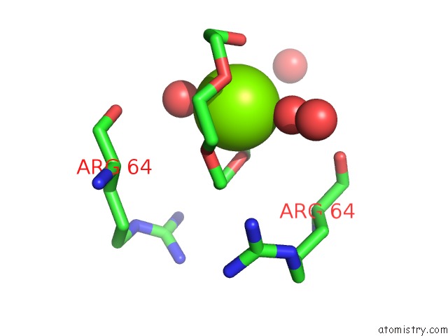
Magnesium binding site 1 out of 1 in 4ymg
Go back to
Magnesium binding site 1 out
of 1 in the Crystal Structure of Sam-Bound Podospora Anserina Methyltransferase PAMTH1
Mono view

Stereo pair view
Mono view
Stereo pair view
A full contact list of Magnesium with other atoms in the Mg binding
site number 1 of Crystal Structure of Sam-Bound Podospora Anserina Methyltransferase PAMTH1 within 5.0Å range:
|
Reference:
D.Chaterjee,
D.Kudlinzki,
V.L.Linhard,
K.Saxena,
U.Schieborr,
S.L.Gande,
J.P.Wurm,
J.Woenert,
R.Abele,
V.V.Rogov,
V.Deotsch,
H.D.Osiewacz,
S.Sreeramulu,
H.Schwalbe.
Structure and Biophysical Characterization of the S-Adenosylmethionine Dependent O-Methyltransferase PAMTH1, A Putative Enzyme Accumulating During Senescence of Podospora Anserina J.Biol.Chem. 2015.
ISSN: ESSN 1083-351X
DOI: 10.1074/JBC.M115.660829
Page generated: Sat Sep 28 23:22:03 2024
ISSN: ESSN 1083-351X
DOI: 10.1074/JBC.M115.660829
Last articles
Zn in 9J0NZn in 9J0O
Zn in 9J0P
Zn in 9FJX
Zn in 9EKB
Zn in 9C0F
Zn in 9CAH
Zn in 9CH0
Zn in 9CH3
Zn in 9CH1