Magnesium »
PDB 4z4g-4zhq »
4zex »
Magnesium in PDB 4zex: Crystal Structure of PFHAD1 in Complex with Glyceraldehyde-3-Phosphate
Protein crystallography data
The structure of Crystal Structure of PFHAD1 in Complex with Glyceraldehyde-3-Phosphate, PDB code: 4zex
was solved by
J.Park,
N.H.Tolia,
with X-Ray Crystallography technique. A brief refinement statistics is given in the table below:
| Resolution Low / High (Å) | 19.93 / 2.00 |
| Space group | P 1 21 1 |
| Cell size a, b, c (Å), α, β, γ (°) | 77.600, 44.600, 84.100, 90.00, 101.30, 90.00 |
| R / Rfree (%) | 17.3 / 22.1 |
Magnesium Binding Sites:
The binding sites of Magnesium atom in the Crystal Structure of PFHAD1 in Complex with Glyceraldehyde-3-Phosphate
(pdb code 4zex). This binding sites where shown within
5.0 Angstroms radius around Magnesium atom.
In total 2 binding sites of Magnesium where determined in the Crystal Structure of PFHAD1 in Complex with Glyceraldehyde-3-Phosphate, PDB code: 4zex:
Jump to Magnesium binding site number: 1; 2;
In total 2 binding sites of Magnesium where determined in the Crystal Structure of PFHAD1 in Complex with Glyceraldehyde-3-Phosphate, PDB code: 4zex:
Jump to Magnesium binding site number: 1; 2;
Magnesium binding site 1 out of 2 in 4zex
Go back to
Magnesium binding site 1 out
of 2 in the Crystal Structure of PFHAD1 in Complex with Glyceraldehyde-3-Phosphate
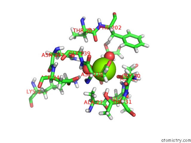
Mono view
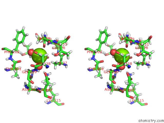
Stereo pair view
Mono view
Stereo pair view
A full contact list of Magnesium with other atoms in the Mg binding
site number 1 of Crystal Structure of PFHAD1 in Complex with Glyceraldehyde-3-Phosphate within 5.0Å range:
|
Magnesium binding site 2 out of 2 in 4zex
Go back to
Magnesium binding site 2 out
of 2 in the Crystal Structure of PFHAD1 in Complex with Glyceraldehyde-3-Phosphate
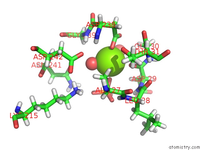
Mono view
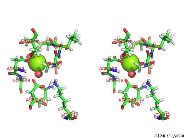
Stereo pair view
Mono view
Stereo pair view
A full contact list of Magnesium with other atoms in the Mg binding
site number 2 of Crystal Structure of PFHAD1 in Complex with Glyceraldehyde-3-Phosphate within 5.0Å range:
|
Reference:
J.Park,
A.M.Guggisberg,
A.R.Odom,
N.H.Tolia.
Cap-Domain Closure Enables Diverse Substrate Recognition By the C2-Type Haloacid Dehalogenase-Like Sugar Phosphatase Plasmodium Falciparum HAD1. Acta Crystallogr. D Biol. V. 71 1824 2015CRYSTALLOGR..
ISSN: ESSN 1399-0047
PubMed: 26327372
DOI: 10.1107/S1399004715012067
Page generated: Sat Sep 28 23:59:49 2024
ISSN: ESSN 1399-0047
PubMed: 26327372
DOI: 10.1107/S1399004715012067
Last articles
Zn in 9JYWZn in 9IR4
Zn in 9IR3
Zn in 9GMX
Zn in 9GMW
Zn in 9JEJ
Zn in 9ERF
Zn in 9ERE
Zn in 9EGV
Zn in 9EGW