Magnesium »
PDB 5ac0-5avx »
5avw »
Magnesium in PDB 5avw: Kinetics By X-Ray Crystallography: Tl+-Substitution of Bound K+ in the E2.MGF42-.2K+ Crystal After 16.5 Min
Protein crystallography data
The structure of Kinetics By X-Ray Crystallography: Tl+-Substitution of Bound K+ in the E2.MGF42-.2K+ Crystal After 16.5 Min, PDB code: 5avw
was solved by
H.Ogawa,
F.Cornelius,
A.Hirata,
C.Toyoshima,
with X-Ray Crystallography technique. A brief refinement statistics is given in the table below:
| Resolution Low / High (Å) | 14.98 / 2.60 |
| Space group | C 1 2 1 |
| Cell size a, b, c (Å), α, β, γ (°) | 222.476, 50.881, 163.810, 90.00, 104.67, 90.00 |
| R / Rfree (%) | 26.5 / 25.7 |
Other elements in 5avw:
The structure of Kinetics By X-Ray Crystallography: Tl+-Substitution of Bound K+ in the E2.MGF42-.2K+ Crystal After 16.5 Min also contains other interesting chemical elements:
| Fluorine | (F) | 4 atoms |
| Potassium | (K) | 1 atom |
| Thallium | (Tl) | 3 atoms |
Magnesium Binding Sites:
The binding sites of Magnesium atom in the Kinetics By X-Ray Crystallography: Tl+-Substitution of Bound K+ in the E2.MGF42-.2K+ Crystal After 16.5 Min
(pdb code 5avw). This binding sites where shown within
5.0 Angstroms radius around Magnesium atom.
In total 2 binding sites of Magnesium where determined in the Kinetics By X-Ray Crystallography: Tl+-Substitution of Bound K+ in the E2.MGF42-.2K+ Crystal After 16.5 Min, PDB code: 5avw:
Jump to Magnesium binding site number: 1; 2;
In total 2 binding sites of Magnesium where determined in the Kinetics By X-Ray Crystallography: Tl+-Substitution of Bound K+ in the E2.MGF42-.2K+ Crystal After 16.5 Min, PDB code: 5avw:
Jump to Magnesium binding site number: 1; 2;
Magnesium binding site 1 out of 2 in 5avw
Go back to
Magnesium binding site 1 out
of 2 in the Kinetics By X-Ray Crystallography: Tl+-Substitution of Bound K+ in the E2.MGF42-.2K+ Crystal After 16.5 Min
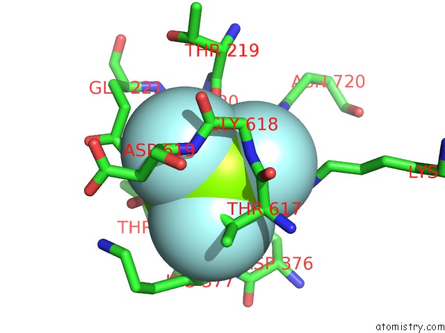
Mono view
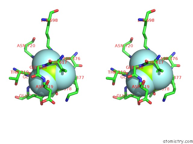
Stereo pair view
Mono view
Stereo pair view
A full contact list of Magnesium with other atoms in the Mg binding
site number 1 of Kinetics By X-Ray Crystallography: Tl+-Substitution of Bound K+ in the E2.MGF42-.2K+ Crystal After 16.5 Min within 5.0Å range:
|
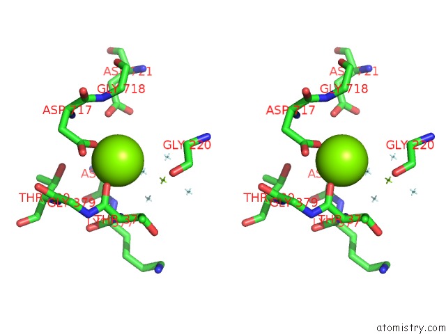
Magnesium binding site 2 out of 2 in 5avw
Go back to
Magnesium binding site 2 out
of 2 in the Kinetics By X-Ray Crystallography: Tl+-Substitution of Bound K+ in the E2.MGF42-.2K+ Crystal After 16.5 Min
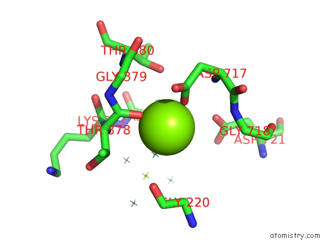
Mono view
Stereo pair view
Mono view
Stereo pair view
A full contact list of Magnesium with other atoms in the Mg binding
site number 2 of Kinetics By X-Ray Crystallography: Tl+-Substitution of Bound K+ in the E2.MGF42-.2K+ Crystal After 16.5 Min within 5.0Å range:
|
Reference:
H.Ogawa,
F.Cornelius,
A.Hirata,
C.Toyoshima.
Sequential Substitution of K(+) Bound to Na(+),K(+)-Atpase Visualized By X-Ray Crystallography. Nat Commun V. 6 8004 2015.
ISSN: ESSN 2041-1723
PubMed: 26258479
DOI: 10.1038/NCOMMS9004
Page generated: Sun Sep 29 00:37:56 2024
ISSN: ESSN 2041-1723
PubMed: 26258479
DOI: 10.1038/NCOMMS9004
Last articles
Zn in 9J0NZn in 9J0O
Zn in 9J0P
Zn in 9FJX
Zn in 9EKB
Zn in 9C0F
Zn in 9CAH
Zn in 9CH0
Zn in 9CH3
Zn in 9CH1