Magnesium »
PDB 5avy-5b8f »
5awm »
Magnesium in PDB 5awm: The Crystal Structure of Jnk From Drosophila Melanogaster Reveals An Evolutionarily Conserved Topology with That of Mammalian Jnk Proteins.
Enzymatic activity of The Crystal Structure of Jnk From Drosophila Melanogaster Reveals An Evolutionarily Conserved Topology with That of Mammalian Jnk Proteins.
All present enzymatic activity of The Crystal Structure of Jnk From Drosophila Melanogaster Reveals An Evolutionarily Conserved Topology with That of Mammalian Jnk Proteins.:
2.7.11.24;
2.7.11.24;
Protein crystallography data
The structure of The Crystal Structure of Jnk From Drosophila Melanogaster Reveals An Evolutionarily Conserved Topology with That of Mammalian Jnk Proteins., PDB code: 5awm
was solved by
P.Boonserm,
with X-Ray Crystallography technique. A brief refinement statistics is given in the table below:
| Resolution Low / High (Å) | 28.28 / 1.79 |
| Space group | P 21 21 21 |
| Cell size a, b, c (Å), α, β, γ (°) | 52.486, 55.329, 126.717, 90.00, 90.00, 90.00 |
| R / Rfree (%) | 17.5 / 22 |
Magnesium Binding Sites:
The binding sites of Magnesium atom in the The Crystal Structure of Jnk From Drosophila Melanogaster Reveals An Evolutionarily Conserved Topology with That of Mammalian Jnk Proteins.
(pdb code 5awm). This binding sites where shown within
5.0 Angstroms radius around Magnesium atom.
In total 2 binding sites of Magnesium where determined in the The Crystal Structure of Jnk From Drosophila Melanogaster Reveals An Evolutionarily Conserved Topology with That of Mammalian Jnk Proteins., PDB code: 5awm:
Jump to Magnesium binding site number: 1; 2;
In total 2 binding sites of Magnesium where determined in the The Crystal Structure of Jnk From Drosophila Melanogaster Reveals An Evolutionarily Conserved Topology with That of Mammalian Jnk Proteins., PDB code: 5awm:
Jump to Magnesium binding site number: 1; 2;
Magnesium binding site 1 out of 2 in 5awm
Go back to
Magnesium binding site 1 out
of 2 in the The Crystal Structure of Jnk From Drosophila Melanogaster Reveals An Evolutionarily Conserved Topology with That of Mammalian Jnk Proteins.
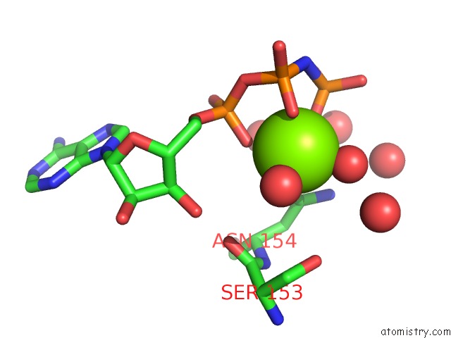
Mono view
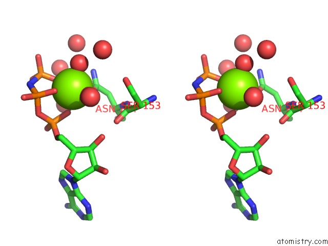
Stereo pair view
Mono view
Stereo pair view
A full contact list of Magnesium with other atoms in the Mg binding
site number 1 of The Crystal Structure of Jnk From Drosophila Melanogaster Reveals An Evolutionarily Conserved Topology with That of Mammalian Jnk Proteins. within 5.0Å range:
|
Magnesium binding site 2 out of 2 in 5awm
Go back to
Magnesium binding site 2 out
of 2 in the The Crystal Structure of Jnk From Drosophila Melanogaster Reveals An Evolutionarily Conserved Topology with That of Mammalian Jnk Proteins.
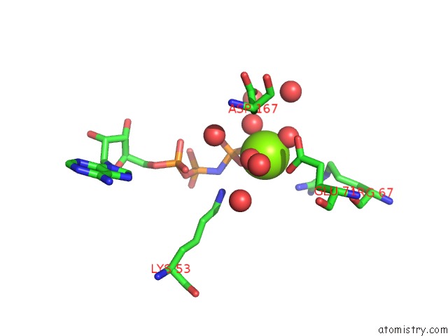
Mono view
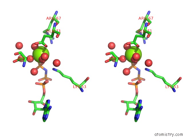
Stereo pair view
Mono view
Stereo pair view
A full contact list of Magnesium with other atoms in the Mg binding
site number 2 of The Crystal Structure of Jnk From Drosophila Melanogaster Reveals An Evolutionarily Conserved Topology with That of Mammalian Jnk Proteins. within 5.0Å range:
|
Reference:
S.Chimnaronk,
J.Sitthiroongruang,
K.Srisucharitpanit,
M.Srisaisup,
A.J.Ketterman,
P.Boonserm.
The Crystal Structure of Jnk From Drosophila Melanogaster Reveals An Evolutionarily Conserved Topology with That of Mammalian Jnk Proteins. Bmc Struct.Biol. V. 15 17 2015.
ISSN: ESSN 1472-6807
PubMed: 26377800
DOI: 10.1186/S12900-015-0045-1
Page generated: Sun Sep 29 00:56:55 2024
ISSN: ESSN 1472-6807
PubMed: 26377800
DOI: 10.1186/S12900-015-0045-1
Last articles
Zn in 9J0NZn in 9J0O
Zn in 9J0P
Zn in 9FJX
Zn in 9EKB
Zn in 9C0F
Zn in 9CAH
Zn in 9CH0
Zn in 9CH3
Zn in 9CH1