Magnesium »
PDB 5bjp-5btm »
5bof »
Magnesium in PDB 5bof: Crystal Structure of Staphylococcus Aureus Enolase
Enzymatic activity of Crystal Structure of Staphylococcus Aureus Enolase
All present enzymatic activity of Crystal Structure of Staphylococcus Aureus Enolase:
4.2.1.11;
4.2.1.11;
Protein crystallography data
The structure of Crystal Structure of Staphylococcus Aureus Enolase, PDB code: 5bof
was solved by
Y.F.Wu,
C.L.Wang,
M.H.Wu,
L.Han,
X.Zhang,
J.Y.Zang,
with X-Ray Crystallography technique. A brief refinement statistics is given in the table below:
| Resolution Low / High (Å) | 50.00 / 2.45 |
| Space group | P 4 21 2 |
| Cell size a, b, c (Å), α, β, γ (°) | 164.686, 164.686, 77.335, 90.00, 90.00, 90.00 |
| R / Rfree (%) | 18.6 / 22.9 |
Magnesium Binding Sites:
The binding sites of Magnesium atom in the Crystal Structure of Staphylococcus Aureus Enolase
(pdb code 5bof). This binding sites where shown within
5.0 Angstroms radius around Magnesium atom.
In total 2 binding sites of Magnesium where determined in the Crystal Structure of Staphylococcus Aureus Enolase, PDB code: 5bof:
Jump to Magnesium binding site number: 1; 2;
In total 2 binding sites of Magnesium where determined in the Crystal Structure of Staphylococcus Aureus Enolase, PDB code: 5bof:
Jump to Magnesium binding site number: 1; 2;
Magnesium binding site 1 out of 2 in 5bof
Go back to
Magnesium binding site 1 out
of 2 in the Crystal Structure of Staphylococcus Aureus Enolase
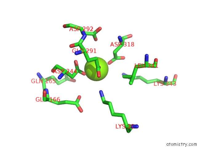
Mono view
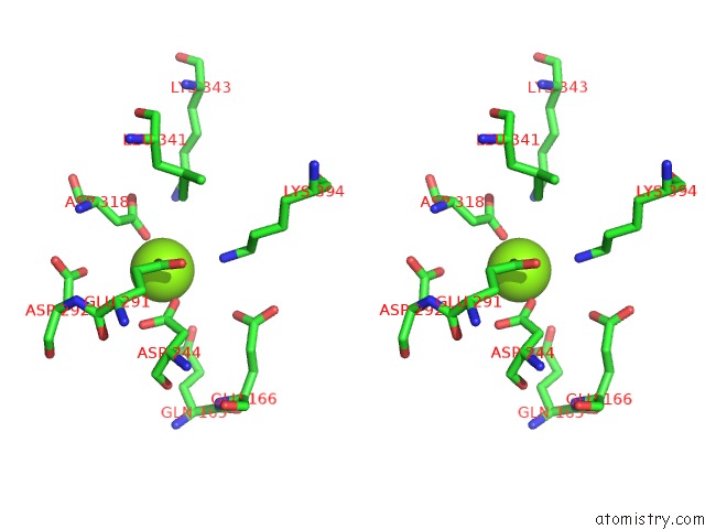
Stereo pair view
Mono view
Stereo pair view
A full contact list of Magnesium with other atoms in the Mg binding
site number 1 of Crystal Structure of Staphylococcus Aureus Enolase within 5.0Å range:
|
Magnesium binding site 2 out of 2 in 5bof
Go back to
Magnesium binding site 2 out
of 2 in the Crystal Structure of Staphylococcus Aureus Enolase
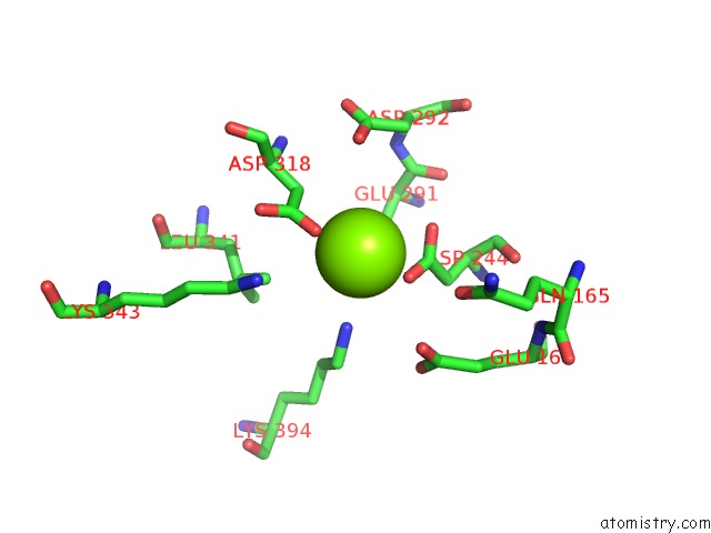
Mono view
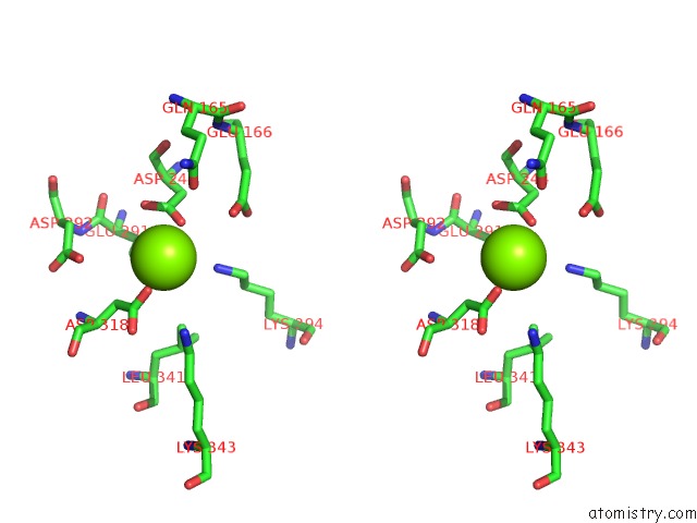
Stereo pair view
Mono view
Stereo pair view
A full contact list of Magnesium with other atoms in the Mg binding
site number 2 of Crystal Structure of Staphylococcus Aureus Enolase within 5.0Å range:
|
Reference:
Y.Wu,
C.Wang,
S.Lin,
M.Wu,
L.Han,
C.Tian,
X.Zhang,
J.Zang.
Octameric Structure of Staphylococcus Aureus Enolase in Complex with Phosphoenolpyruvate. Acta Crystallogr.,Sect.D V. 71 2457 2015.
ISSN: ESSN 1399-0047
PubMed: 26627653
DOI: 10.1107/S1399004715018830
Page generated: Sun Sep 29 01:12:59 2024
ISSN: ESSN 1399-0047
PubMed: 26627653
DOI: 10.1107/S1399004715018830
Last articles
Fe in 2YXOFe in 2YRS
Fe in 2YXC
Fe in 2YNM
Fe in 2YVJ
Fe in 2YP1
Fe in 2YU2
Fe in 2YU1
Fe in 2YQB
Fe in 2YOO