Magnesium »
PDB 5bjp-5btm »
5bpj »
Magnesium in PDB 5bpj: All Three Ca(2+)-Binding Loops of Light-Sensitive Ctenophore Photoprotein Berovin Bind Magnesium Ions: the Spatial Structure of Mg(2+)-Loaded Apo-Berovin
Protein crystallography data
The structure of All Three Ca(2+)-Binding Loops of Light-Sensitive Ctenophore Photoprotein Berovin Bind Magnesium Ions: the Spatial Structure of Mg(2+)-Loaded Apo-Berovin, PDB code: 5bpj
was solved by
L.P.Burakova,
P.V.Natashin,
N.P.Malikova,
F.Niu,
E.S.Vysotski,
Z.-J.Liu,
with X-Ray Crystallography technique. A brief refinement statistics is given in the table below:
| Resolution Low / High (Å) | 24.59 / 1.76 |
| Space group | C 1 2 1 |
| Cell size a, b, c (Å), α, β, γ (°) | 94.816, 33.165, 72.074, 90.00, 126.70, 90.00 |
| R / Rfree (%) | 22.2 / 24.3 |
Magnesium Binding Sites:
The binding sites of Magnesium atom in the All Three Ca(2+)-Binding Loops of Light-Sensitive Ctenophore Photoprotein Berovin Bind Magnesium Ions: the Spatial Structure of Mg(2+)-Loaded Apo-Berovin
(pdb code 5bpj). This binding sites where shown within
5.0 Angstroms radius around Magnesium atom.
In total 3 binding sites of Magnesium where determined in the All Three Ca(2+)-Binding Loops of Light-Sensitive Ctenophore Photoprotein Berovin Bind Magnesium Ions: the Spatial Structure of Mg(2+)-Loaded Apo-Berovin, PDB code: 5bpj:
Jump to Magnesium binding site number: 1; 2; 3;
In total 3 binding sites of Magnesium where determined in the All Three Ca(2+)-Binding Loops of Light-Sensitive Ctenophore Photoprotein Berovin Bind Magnesium Ions: the Spatial Structure of Mg(2+)-Loaded Apo-Berovin, PDB code: 5bpj:
Jump to Magnesium binding site number: 1; 2; 3;
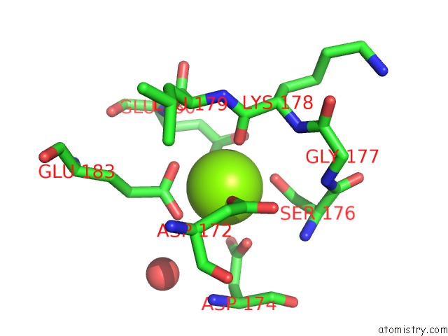
Magnesium binding site 1 out of 3 in 5bpj
Go back to
Magnesium binding site 1 out
of 3 in the All Three Ca(2+)-Binding Loops of Light-Sensitive Ctenophore Photoprotein Berovin Bind Magnesium Ions: the Spatial Structure of Mg(2+)-Loaded Apo-Berovin
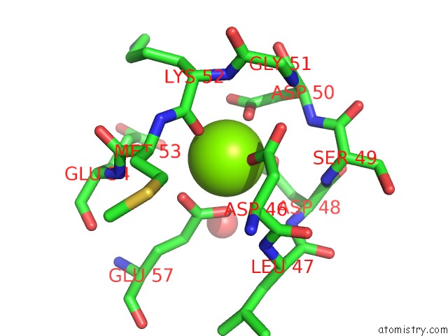
Mono view
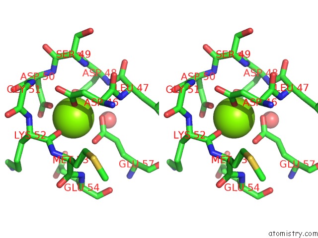
Stereo pair view
Mono view
Stereo pair view
A full contact list of Magnesium with other atoms in the Mg binding
site number 1 of All Three Ca(2+)-Binding Loops of Light-Sensitive Ctenophore Photoprotein Berovin Bind Magnesium Ions: the Spatial Structure of Mg(2+)-Loaded Apo-Berovin within 5.0Å range:
|
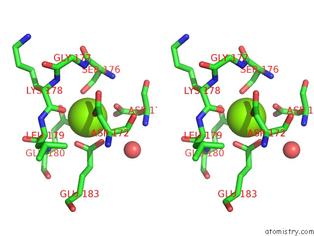
Magnesium binding site 2 out of 3 in 5bpj
Go back to
Magnesium binding site 2 out
of 3 in the All Three Ca(2+)-Binding Loops of Light-Sensitive Ctenophore Photoprotein Berovin Bind Magnesium Ions: the Spatial Structure of Mg(2+)-Loaded Apo-Berovin
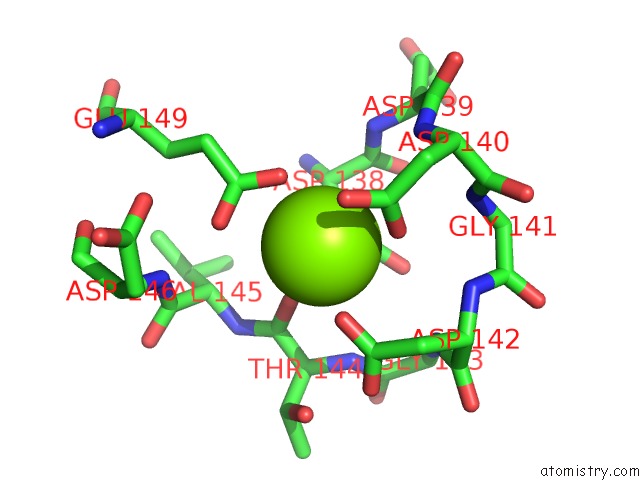
Mono view
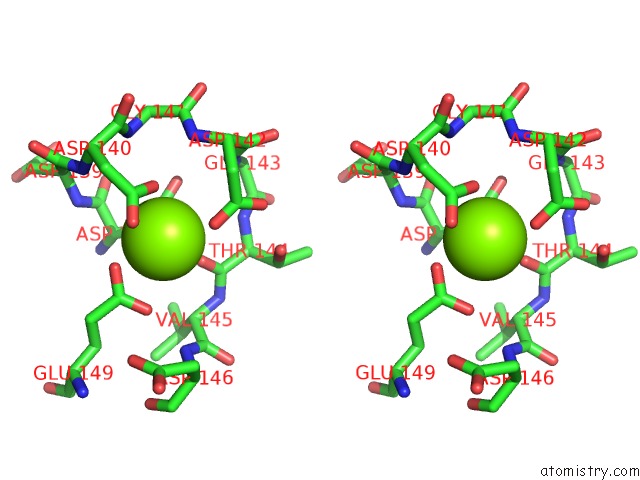
Stereo pair view
Mono view
Stereo pair view
A full contact list of Magnesium with other atoms in the Mg binding
site number 2 of All Three Ca(2+)-Binding Loops of Light-Sensitive Ctenophore Photoprotein Berovin Bind Magnesium Ions: the Spatial Structure of Mg(2+)-Loaded Apo-Berovin within 5.0Å range:
|
Magnesium binding site 3 out of 3 in 5bpj
Go back to
Magnesium binding site 3 out
of 3 in the All Three Ca(2+)-Binding Loops of Light-Sensitive Ctenophore Photoprotein Berovin Bind Magnesium Ions: the Spatial Structure of Mg(2+)-Loaded Apo-Berovin
Mono view
Stereo pair view
Mono view
Stereo pair view
A full contact list of Magnesium with other atoms in the Mg binding
site number 3 of All Three Ca(2+)-Binding Loops of Light-Sensitive Ctenophore Photoprotein Berovin Bind Magnesium Ions: the Spatial Structure of Mg(2+)-Loaded Apo-Berovin within 5.0Å range:
|
Reference:
L.P.Burakova,
P.V.Natashin,
N.P.Malikova,
F.Niu,
M.Pu,
E.S.Vysotski,
Z.J.Liu.
All Ca(2+)-Binding Loops of Light-Sensitive Ctenophore Photoprotein Berovin Bind Magnesium Ions: the Spatial Structure of Mg(2+)-Loaded Apo-Berovin. J. Photochem. Photobiol. B, V. 154 57 2016BIOL..
ISSN: ISSN 1873-2682
PubMed: 26690016
DOI: 10.1016/J.JPHOTOBIOL.2015.11.012
Page generated: Tue Aug 12 05:43:10 2025
ISSN: ISSN 1873-2682
PubMed: 26690016
DOI: 10.1016/J.JPHOTOBIOL.2015.11.012
Last articles
Mg in 5GG8Mg in 5GG7
Mg in 5GG6
Mg in 5GAD
Mg in 5GAG
Mg in 5GAF
Mg in 5GAE
Mg in 5G0R
Mg in 5G5V
Mg in 5G3T