Magnesium »
PDB 5bjp-5btm »
5bst »
Magnesium in PDB 5bst: Crystal Structure of 4-Coumarate:Coa Ligase Complexed with Coumaroyl Adenylate
Enzymatic activity of Crystal Structure of 4-Coumarate:Coa Ligase Complexed with Coumaroyl Adenylate
All present enzymatic activity of Crystal Structure of 4-Coumarate:Coa Ligase Complexed with Coumaroyl Adenylate:
6.2.1.12;
6.2.1.12;
Protein crystallography data
The structure of Crystal Structure of 4-Coumarate:Coa Ligase Complexed with Coumaroyl Adenylate, PDB code: 5bst
was solved by
Z.Li,
S.K.Nair,
with X-Ray Crystallography technique. A brief refinement statistics is given in the table below:
| Resolution Low / High (Å) | 25.00 / 1.61 |
| Space group | P 41 21 2 |
| Cell size a, b, c (Å), α, β, γ (°) | 81.830, 81.830, 180.988, 90.00, 90.00, 90.00 |
| R / Rfree (%) | 19.9 / 21.5 |
Magnesium Binding Sites:
The binding sites of Magnesium atom in the Crystal Structure of 4-Coumarate:Coa Ligase Complexed with Coumaroyl Adenylate
(pdb code 5bst). This binding sites where shown within
5.0 Angstroms radius around Magnesium atom.
In total only one binding site of Magnesium was determined in the Crystal Structure of 4-Coumarate:Coa Ligase Complexed with Coumaroyl Adenylate, PDB code: 5bst:
In total only one binding site of Magnesium was determined in the Crystal Structure of 4-Coumarate:Coa Ligase Complexed with Coumaroyl Adenylate, PDB code: 5bst:
Magnesium binding site 1 out of 1 in 5bst
Go back to
Magnesium binding site 1 out
of 1 in the Crystal Structure of 4-Coumarate:Coa Ligase Complexed with Coumaroyl Adenylate
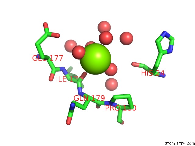
Mono view
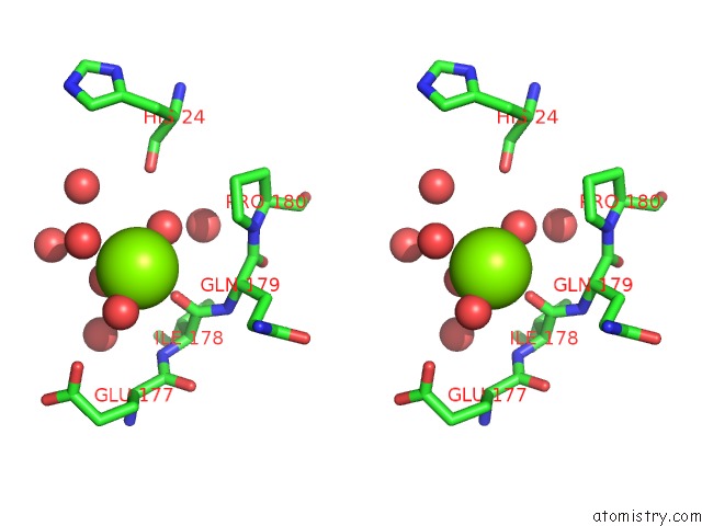
Stereo pair view
Mono view
Stereo pair view
A full contact list of Magnesium with other atoms in the Mg binding
site number 1 of Crystal Structure of 4-Coumarate:Coa Ligase Complexed with Coumaroyl Adenylate within 5.0Å range:
|
Reference:
Z.Li,
S.K.Nair.
Structural Basis For Specificity and Flexibility in A Plant 4-Coumarate:Coa Ligase. Structure V. 23 2032 2015.
ISSN: ISSN 0969-2126
PubMed: 26412334
DOI: 10.1016/J.STR.2015.08.012
Page generated: Tue Aug 12 05:48:11 2025
ISSN: ISSN 0969-2126
PubMed: 26412334
DOI: 10.1016/J.STR.2015.08.012
Last articles
Mg in 5GG8Mg in 5GG7
Mg in 5GG6
Mg in 5GAD
Mg in 5GAG
Mg in 5GAF
Mg in 5GAE
Mg in 5G0R
Mg in 5G5V
Mg in 5G3T