Magnesium »
PDB 5btn-5c28 »
5byg »
Magnesium in PDB 5byg: X-Ray Structure of AAV2 Obd-AAVS1 Complex 2:1
Protein crystallography data
The structure of X-Ray Structure of AAV2 Obd-AAVS1 Complex 2:1, PDB code: 5byg
was solved by
F.N.Musayev,
C.R.Escalante,
with X-Ray Crystallography technique. A brief refinement statistics is given in the table below:
| Resolution Low / High (Å) | 29.40 / 2.50 |
| Space group | C 1 2 1 |
| Cell size a, b, c (Å), α, β, γ (°) | 183.136, 79.595, 139.502, 90.00, 98.12, 90.00 |
| R / Rfree (%) | 18.9 / 22.7 |
Magnesium Binding Sites:
The binding sites of Magnesium atom in the X-Ray Structure of AAV2 Obd-AAVS1 Complex 2:1
(pdb code 5byg). This binding sites where shown within
5.0 Angstroms radius around Magnesium atom.
In total 3 binding sites of Magnesium where determined in the X-Ray Structure of AAV2 Obd-AAVS1 Complex 2:1, PDB code: 5byg:
Jump to Magnesium binding site number: 1; 2; 3;
In total 3 binding sites of Magnesium where determined in the X-Ray Structure of AAV2 Obd-AAVS1 Complex 2:1, PDB code: 5byg:
Jump to Magnesium binding site number: 1; 2; 3;
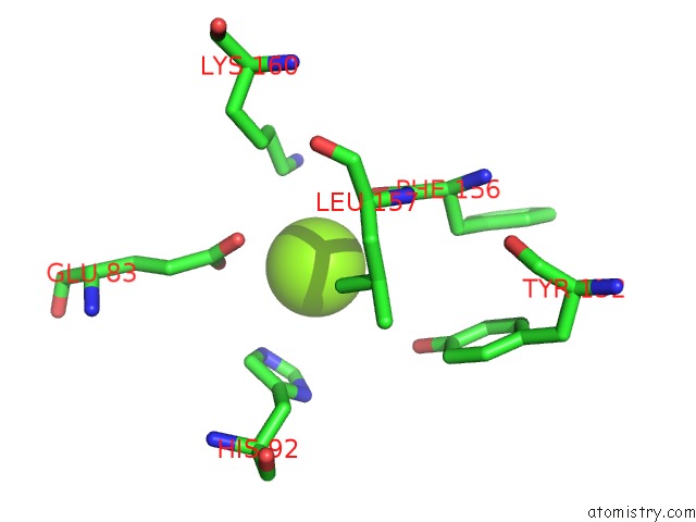
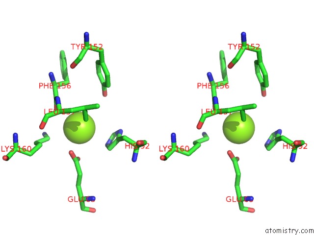
Magnesium binding site 1 out of 3 in 5byg
Go back to
Magnesium binding site 1 out
of 3 in the X-Ray Structure of AAV2 Obd-AAVS1 Complex 2:1
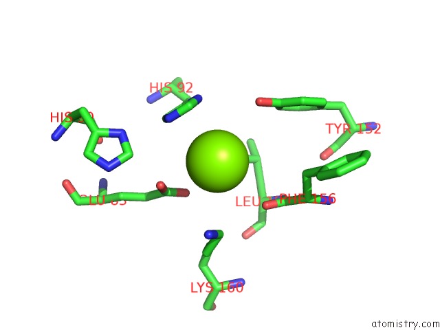
Mono view
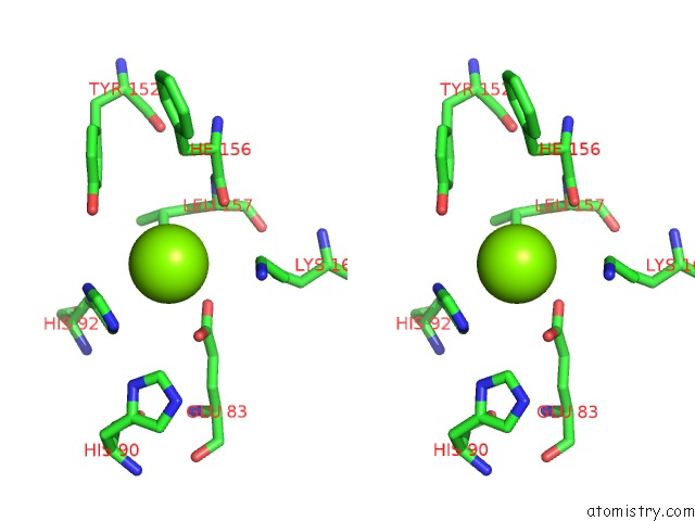
Stereo pair view
Mono view
Stereo pair view
A full contact list of Magnesium with other atoms in the Mg binding
site number 1 of X-Ray Structure of AAV2 Obd-AAVS1 Complex 2:1 within 5.0Å range:
|
Magnesium binding site 2 out of 3 in 5byg
Go back to
Magnesium binding site 2 out
of 3 in the X-Ray Structure of AAV2 Obd-AAVS1 Complex 2:1
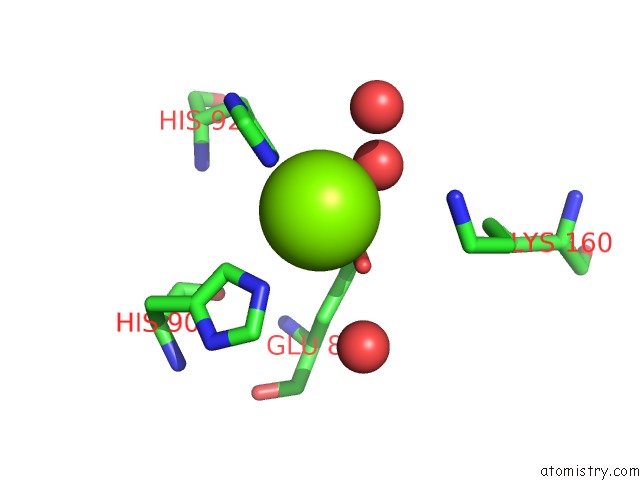
Mono view
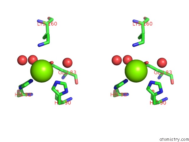
Stereo pair view
Mono view
Stereo pair view
A full contact list of Magnesium with other atoms in the Mg binding
site number 2 of X-Ray Structure of AAV2 Obd-AAVS1 Complex 2:1 within 5.0Å range:
|
Magnesium binding site 3 out of 3 in 5byg
Go back to
Magnesium binding site 3 out
of 3 in the X-Ray Structure of AAV2 Obd-AAVS1 Complex 2:1
Mono view
Stereo pair view
Mono view
Stereo pair view
A full contact list of Magnesium with other atoms in the Mg binding
site number 3 of X-Ray Structure of AAV2 Obd-AAVS1 Complex 2:1 within 5.0Å range:
|
Reference:
F.N.Musayev,
F.Zarate-Perez,
C.Bishop,
J.W.Burgner,
C.R.Escalante.
Structural Insights Into the Assembly of the Adeno-Associated Virus Type 2 REP68 Protein on the Integration Site AAVS1. J.Biol.Chem. V. 290 27487 2015.
ISSN: ESSN 1083-351X
PubMed: 26370092
DOI: 10.1074/JBC.M115.669960
Page generated: Sun Sep 29 01:47:08 2024
ISSN: ESSN 1083-351X
PubMed: 26370092
DOI: 10.1074/JBC.M115.669960
Last articles
Zn in 9MJ5Zn in 9HNW
Zn in 9G0L
Zn in 9FNE
Zn in 9DZN
Zn in 9E0I
Zn in 9D32
Zn in 9DAK
Zn in 8ZXC
Zn in 8ZUF