Magnesium »
PDB 5btn-5c28 »
5bzy »
Magnesium in PDB 5bzy: X-Ray Crystal Structure of A Continuously Hydrogen Bonded 14MER Dna Lattice.
Protein crystallography data
The structure of X-Ray Crystal Structure of A Continuously Hydrogen Bonded 14MER Dna Lattice., PDB code: 5bzy
was solved by
M.Saoji,
P.J.Paukstelis,
with X-Ray Crystallography technique. A brief refinement statistics is given in the table below:
| Resolution Low / High (Å) | 40.51 / 2.40 |
| Space group | P 31 2 1 |
| Cell size a, b, c (Å), α, β, γ (°) | 25.958, 25.958, 121.533, 90.00, 90.00, 120.00 |
| R / Rfree (%) | 24.5 / 31.3 |
Magnesium Binding Sites:
The binding sites of Magnesium atom in the X-Ray Crystal Structure of A Continuously Hydrogen Bonded 14MER Dna Lattice.
(pdb code 5bzy). This binding sites where shown within
5.0 Angstroms radius around Magnesium atom.
In total 2 binding sites of Magnesium where determined in the X-Ray Crystal Structure of A Continuously Hydrogen Bonded 14MER Dna Lattice., PDB code: 5bzy:
Jump to Magnesium binding site number: 1; 2;
In total 2 binding sites of Magnesium where determined in the X-Ray Crystal Structure of A Continuously Hydrogen Bonded 14MER Dna Lattice., PDB code: 5bzy:
Jump to Magnesium binding site number: 1; 2;
Magnesium binding site 1 out of 2 in 5bzy
Go back to
Magnesium binding site 1 out
of 2 in the X-Ray Crystal Structure of A Continuously Hydrogen Bonded 14MER Dna Lattice.
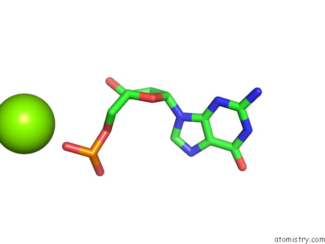
Mono view
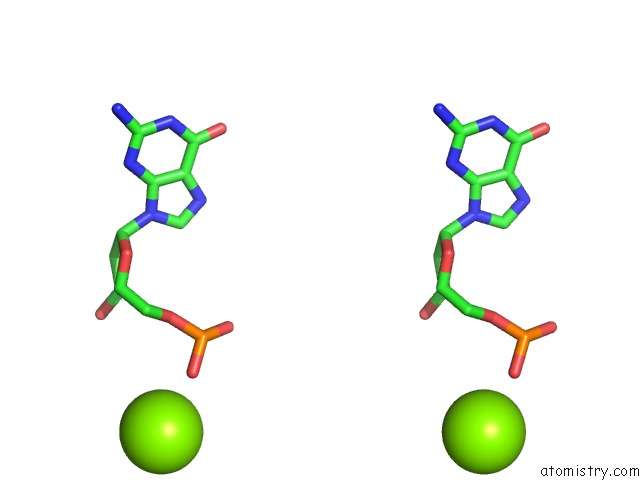
Stereo pair view
Mono view
Stereo pair view
A full contact list of Magnesium with other atoms in the Mg binding
site number 1 of X-Ray Crystal Structure of A Continuously Hydrogen Bonded 14MER Dna Lattice. within 5.0Å range:
|
Magnesium binding site 2 out of 2 in 5bzy
Go back to
Magnesium binding site 2 out
of 2 in the X-Ray Crystal Structure of A Continuously Hydrogen Bonded 14MER Dna Lattice.
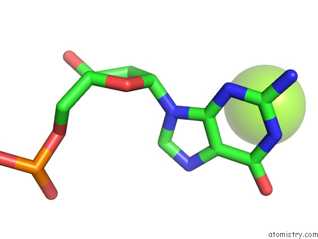
Mono view
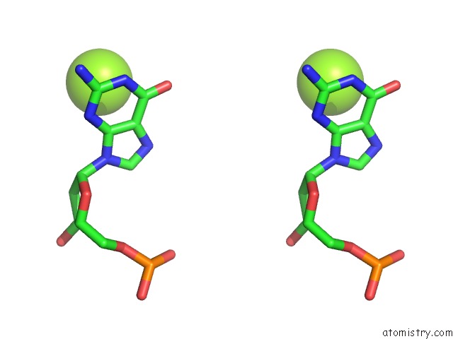
Stereo pair view
Mono view
Stereo pair view
A full contact list of Magnesium with other atoms in the Mg binding
site number 2 of X-Ray Crystal Structure of A Continuously Hydrogen Bonded 14MER Dna Lattice. within 5.0Å range:
|
Reference:
M.Saoji,
P.J.Paukstelis.
Sequence-Dependent Structural Changes in A Self-Assembling Dna Oligonucleotide. Acta Crystallogr.,Sect.D V. 71 2471 2015.
ISSN: ESSN 1399-0047
PubMed: 26627654
DOI: 10.1107/S1399004715019598
Page generated: Sun Sep 29 01:51:00 2024
ISSN: ESSN 1399-0047
PubMed: 26627654
DOI: 10.1107/S1399004715019598
Last articles
Zn in 9MJ5Zn in 9HNW
Zn in 9G0L
Zn in 9FNE
Zn in 9DZN
Zn in 9E0I
Zn in 9D32
Zn in 9DAK
Zn in 8ZXC
Zn in 8ZUF