Magnesium »
PDB 5c28-5ca0 »
5c6t »
Magnesium in PDB 5c6t: Crystal Structure of Hcmv Glycoprotein B in Complex with 1G2 Fab
Protein crystallography data
The structure of Crystal Structure of Hcmv Glycoprotein B in Complex with 1G2 Fab, PDB code: 5c6t
was solved by
S.Chandramouli,
C.Ciferri,
E.C.Settembre,
A.Carfi,
with X-Ray Crystallography technique. A brief refinement statistics is given in the table below:
| Resolution Low / High (Å) | 19.98 / 3.60 |
| Space group | P 21 3 |
| Cell size a, b, c (Å), α, β, γ (°) | 176.486, 176.486, 176.486, 90.00, 90.00, 90.00 |
| R / Rfree (%) | 21.2 / 26 |
Magnesium Binding Sites:
The binding sites of Magnesium atom in the Crystal Structure of Hcmv Glycoprotein B in Complex with 1G2 Fab
(pdb code 5c6t). This binding sites where shown within
5.0 Angstroms radius around Magnesium atom.
In total only one binding site of Magnesium was determined in the Crystal Structure of Hcmv Glycoprotein B in Complex with 1G2 Fab, PDB code: 5c6t:
In total only one binding site of Magnesium was determined in the Crystal Structure of Hcmv Glycoprotein B in Complex with 1G2 Fab, PDB code: 5c6t:
Magnesium binding site 1 out of 1 in 5c6t
Go back to
Magnesium binding site 1 out
of 1 in the Crystal Structure of Hcmv Glycoprotein B in Complex with 1G2 Fab
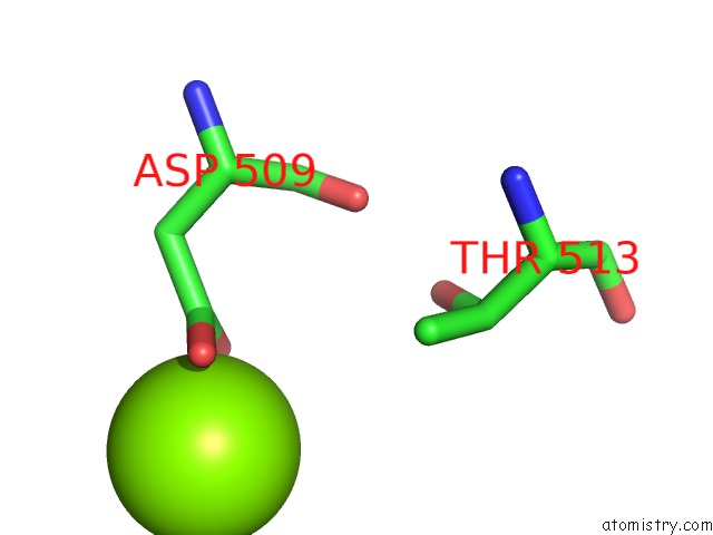
Mono view
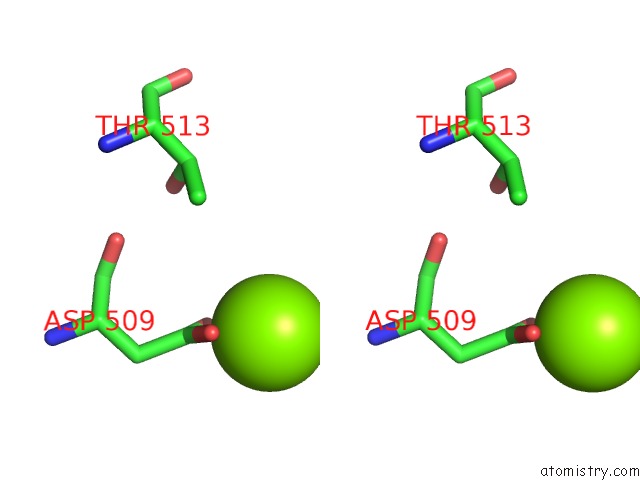
Stereo pair view
Mono view
Stereo pair view
A full contact list of Magnesium with other atoms in the Mg binding
site number 1 of Crystal Structure of Hcmv Glycoprotein B in Complex with 1G2 Fab within 5.0Å range:
|
Reference:
S.Chandramouli,
C.Ciferri,
P.A.Nikitin,
S.Calo,
R.Gerrein,
K.Balabanis,
J.Monroe,
C.Hebner,
A.E.Lilja,
E.C.Settembre,
A.Carfi.
Structure of Hcmv Glycoprotein B in the Postfusion Conformation Bound to A Neutralizing Human Antibody. Nat Commun V. 6 8176 2015.
ISSN: ESSN 2041-1723
PubMed: 26365435
DOI: 10.1038/NCOMMS9176
Page generated: Sun Sep 29 01:58:52 2024
ISSN: ESSN 2041-1723
PubMed: 26365435
DOI: 10.1038/NCOMMS9176
Last articles
Zn in 9J0NZn in 9J0O
Zn in 9J0P
Zn in 9FJX
Zn in 9EKB
Zn in 9C0F
Zn in 9CAH
Zn in 9CH0
Zn in 9CH3
Zn in 9CH1