Magnesium »
PDB 5ca1-5cgi »
5ca3 »
Magnesium in PDB 5ca3: Crystal Structure of the Glycosynthase Mutant D324N of Escherichia Coli GH63 Glycosidase in Complex with Glucose and Lactose
Protein crystallography data
The structure of Crystal Structure of the Glycosynthase Mutant D324N of Escherichia Coli GH63 Glycosidase in Complex with Glucose and Lactose, PDB code: 5ca3
was solved by
T.Miyazaki,
T.Tonozuka,
with X-Ray Crystallography technique. A brief refinement statistics is given in the table below:
| Resolution Low / High (Å) | 31.12 / 1.80 |
| Space group | P 1 21 1 |
| Cell size a, b, c (Å), α, β, γ (°) | 57.423, 136.911, 81.518, 90.00, 100.72, 90.00 |
| R / Rfree (%) | 14.9 / 18.7 |
Other elements in 5ca3:
The structure of Crystal Structure of the Glycosynthase Mutant D324N of Escherichia Coli GH63 Glycosidase in Complex with Glucose and Lactose also contains other interesting chemical elements:
| Calcium | (Ca) | 2 atoms |
Magnesium Binding Sites:
The binding sites of Magnesium atom in the Crystal Structure of the Glycosynthase Mutant D324N of Escherichia Coli GH63 Glycosidase in Complex with Glucose and Lactose
(pdb code 5ca3). This binding sites where shown within
5.0 Angstroms radius around Magnesium atom.
In total 3 binding sites of Magnesium where determined in the Crystal Structure of the Glycosynthase Mutant D324N of Escherichia Coli GH63 Glycosidase in Complex with Glucose and Lactose, PDB code: 5ca3:
Jump to Magnesium binding site number: 1; 2; 3;
In total 3 binding sites of Magnesium where determined in the Crystal Structure of the Glycosynthase Mutant D324N of Escherichia Coli GH63 Glycosidase in Complex with Glucose and Lactose, PDB code: 5ca3:
Jump to Magnesium binding site number: 1; 2; 3;
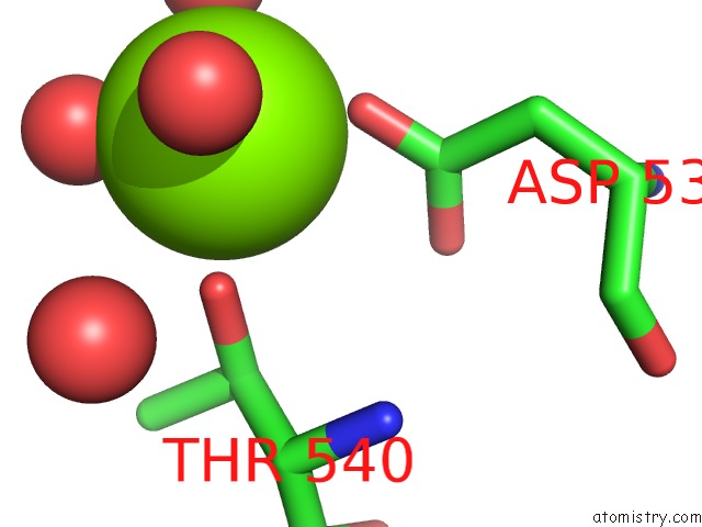
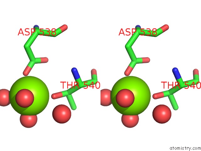
Magnesium binding site 1 out of 3 in 5ca3
Go back to
Magnesium binding site 1 out
of 3 in the Crystal Structure of the Glycosynthase Mutant D324N of Escherichia Coli GH63 Glycosidase in Complex with Glucose and Lactose
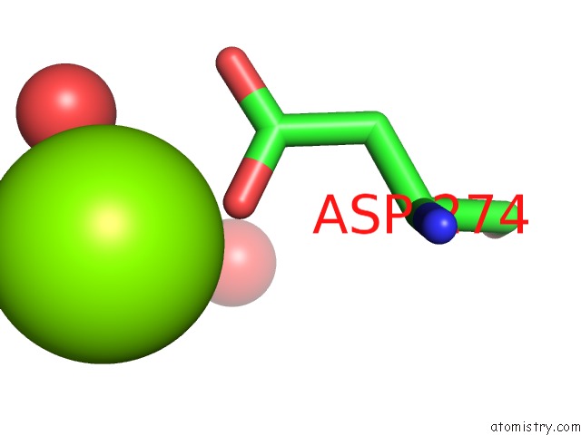
Mono view
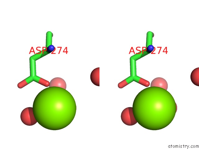
Stereo pair view
Mono view
Stereo pair view
A full contact list of Magnesium with other atoms in the Mg binding
site number 1 of Crystal Structure of the Glycosynthase Mutant D324N of Escherichia Coli GH63 Glycosidase in Complex with Glucose and Lactose within 5.0Å range:
|
Magnesium binding site 2 out of 3 in 5ca3
Go back to
Magnesium binding site 2 out
of 3 in the Crystal Structure of the Glycosynthase Mutant D324N of Escherichia Coli GH63 Glycosidase in Complex with Glucose and Lactose
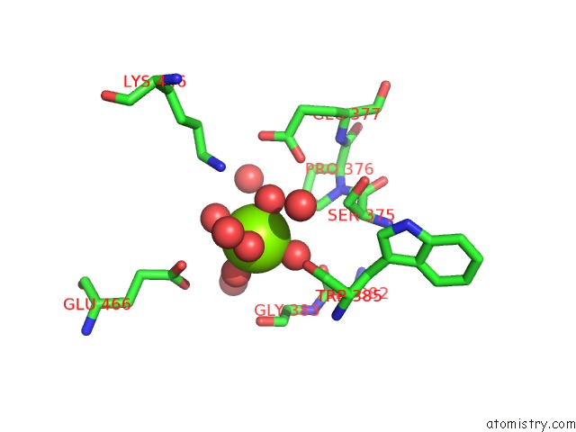
Mono view
Stereo pair view
Mono view
Stereo pair view
A full contact list of Magnesium with other atoms in the Mg binding
site number 2 of Crystal Structure of the Glycosynthase Mutant D324N of Escherichia Coli GH63 Glycosidase in Complex with Glucose and Lactose within 5.0Å range:
|
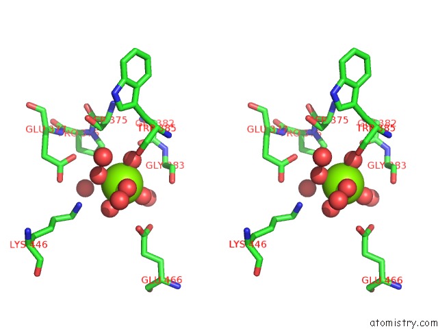
Magnesium binding site 3 out of 3 in 5ca3
Go back to
Magnesium binding site 3 out
of 3 in the Crystal Structure of the Glycosynthase Mutant D324N of Escherichia Coli GH63 Glycosidase in Complex with Glucose and Lactose
Mono view
Stereo pair view
Mono view
Stereo pair view
A full contact list of Magnesium with other atoms in the Mg binding
site number 3 of Crystal Structure of the Glycosynthase Mutant D324N of Escherichia Coli GH63 Glycosidase in Complex with Glucose and Lactose within 5.0Å range:
|
Reference:
T.Miyazaki,
A.Nishikawa,
T.Tonozuka.
Crystal Structure of the Enzyme-Product Complex Reveals Sugar Ring Distortion During Catalysis By Family 63 Inverting Alpha-Glycosidase. J.Struct.Biol. 2016.
ISSN: ESSN 1095-8657
PubMed: 27688023
DOI: 10.1016/J.JSB.2016.09.015
Page generated: Sun Sep 29 02:01:48 2024
ISSN: ESSN 1095-8657
PubMed: 27688023
DOI: 10.1016/J.JSB.2016.09.015
Last articles
Zn in 9J0NZn in 9J0O
Zn in 9J0P
Zn in 9FJX
Zn in 9EKB
Zn in 9C0F
Zn in 9CAH
Zn in 9CH0
Zn in 9CH3
Zn in 9CH1