Magnesium »
PDB 5chi-5cr0 »
5cmb »
Magnesium in PDB 5cmb: Mnemiopsis Leidyi ML032222A Iglur Lbd R703K Mutant Glycine Complex
Protein crystallography data
The structure of Mnemiopsis Leidyi ML032222A Iglur Lbd R703K Mutant Glycine Complex, PDB code: 5cmb
was solved by
M.L.Mayer,
A.Thomas,
with X-Ray Crystallography technique. A brief refinement statistics is given in the table below:
| Resolution Low / High (Å) | 38.99 / 1.34 |
| Space group | P 1 21 1 |
| Cell size a, b, c (Å), α, β, γ (°) | 45.731, 123.389, 54.334, 90.00, 112.20, 90.00 |
| R / Rfree (%) | 14.3 / 16.7 |
Magnesium Binding Sites:
The binding sites of Magnesium atom in the Mnemiopsis Leidyi ML032222A Iglur Lbd R703K Mutant Glycine Complex
(pdb code 5cmb). This binding sites where shown within
5.0 Angstroms radius around Magnesium atom.
In total only one binding site of Magnesium was determined in the Mnemiopsis Leidyi ML032222A Iglur Lbd R703K Mutant Glycine Complex, PDB code: 5cmb:
In total only one binding site of Magnesium was determined in the Mnemiopsis Leidyi ML032222A Iglur Lbd R703K Mutant Glycine Complex, PDB code: 5cmb:
Magnesium binding site 1 out of 1 in 5cmb
Go back to
Magnesium binding site 1 out
of 1 in the Mnemiopsis Leidyi ML032222A Iglur Lbd R703K Mutant Glycine Complex
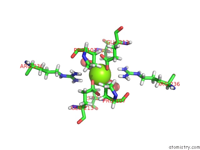
Mono view
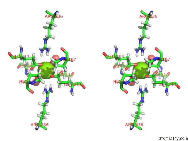
Stereo pair view
Mono view
Stereo pair view
A full contact list of Magnesium with other atoms in the Mg binding
site number 1 of Mnemiopsis Leidyi ML032222A Iglur Lbd R703K Mutant Glycine Complex within 5.0Å range:
|
Reference:
A.Yu,
R.Alberstein,
A.Thomas,
A.Zimmet,
R.Grey,
M.L.Mayer,
A.Y.Lau.
Molecular Lock Regulates Binding of Glycine to A Primitive Nmda Receptor. Proc.Natl.Acad.Sci.Usa V. 113 E6786 2016.
ISSN: ESSN 1091-6490
PubMed: 27791085
DOI: 10.1073/PNAS.1607010113
Page generated: Sun Sep 29 02:14:05 2024
ISSN: ESSN 1091-6490
PubMed: 27791085
DOI: 10.1073/PNAS.1607010113
Last articles
Fe in 2YXOFe in 2YRS
Fe in 2YXC
Fe in 2YNM
Fe in 2YVJ
Fe in 2YP1
Fe in 2YU2
Fe in 2YU1
Fe in 2YQB
Fe in 2YOO