Magnesium »
PDB 5cr0-5d2h »
5cx7 »
Magnesium in PDB 5cx7: Crystal Structure of Pduoc:Heme Complex
Protein crystallography data
The structure of Crystal Structure of Pduoc:Heme Complex, PDB code: 5cx7
was solved by
S.Geremia,
N.Hickey,
D.Ortiz De Orue Lucana,
with X-Ray Crystallography technique. A brief refinement statistics is given in the table below:
| Resolution Low / High (Å) | 33.07 / 1.97 |
| Space group | P 1 21 1 |
| Cell size a, b, c (Å), α, β, γ (°) | 71.300, 130.120, 120.750, 90.00, 90.00, 90.00 |
| R / Rfree (%) | 15.8 / 18.6 |
Other elements in 5cx7:
The structure of Crystal Structure of Pduoc:Heme Complex also contains other interesting chemical elements:
| Iron | (Fe) | 8 atoms |
| Chlorine | (Cl) | 4 atoms |
| Sodium | (Na) | 18 atoms |
Magnesium Binding Sites:
The binding sites of Magnesium atom in the Crystal Structure of Pduoc:Heme Complex
(pdb code 5cx7). This binding sites where shown within
5.0 Angstroms radius around Magnesium atom.
In total 2 binding sites of Magnesium where determined in the Crystal Structure of Pduoc:Heme Complex, PDB code: 5cx7:
Jump to Magnesium binding site number: 1; 2;
In total 2 binding sites of Magnesium where determined in the Crystal Structure of Pduoc:Heme Complex, PDB code: 5cx7:
Jump to Magnesium binding site number: 1; 2;
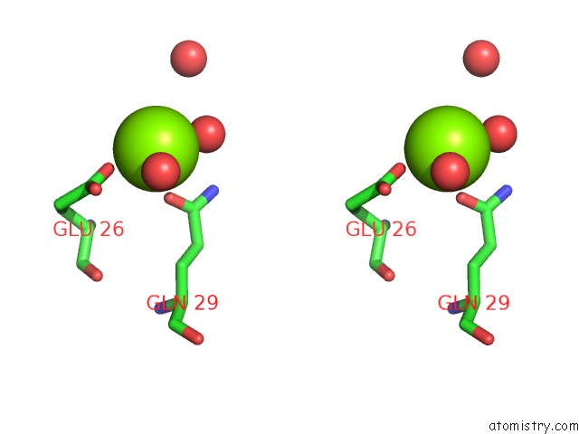
Magnesium binding site 1 out of 2 in 5cx7
Go back to
Magnesium binding site 1 out
of 2 in the Crystal Structure of Pduoc:Heme Complex

Mono view
Stereo pair view
Mono view
Stereo pair view
A full contact list of Magnesium with other atoms in the Mg binding
site number 1 of Crystal Structure of Pduoc:Heme Complex within 5.0Å range:
|
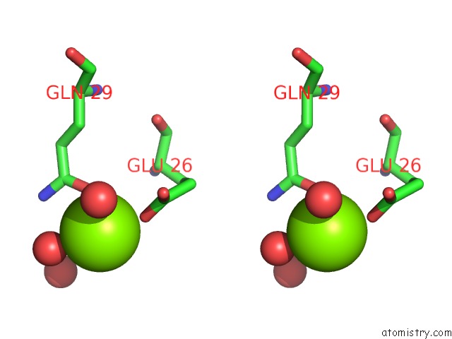
Magnesium binding site 2 out of 2 in 5cx7
Go back to
Magnesium binding site 2 out
of 2 in the Crystal Structure of Pduoc:Heme Complex

Mono view
Stereo pair view
Mono view
Stereo pair view
A full contact list of Magnesium with other atoms in the Mg binding
site number 2 of Crystal Structure of Pduoc:Heme Complex within 5.0Å range:
|
Reference:
D.Ortiz De Orue Lucana,
N.Hickey,
M.Hensel,
J.P.Klare,
S.Geremia,
T.Tiufiakova,
A.E.Torda.
The Crystal Structure of the C-Terminal Domain of the Salmonella Enterica Pduo Protein: An Old Fold with A New Heme-Binding Mode. Front Microbiol V. 7 1010 2016.
ISSN: ESSN 1664-302X
PubMed: 27446048
DOI: 10.3389/FMICB.2016.01010
Page generated: Sun Sep 29 02:19:19 2024
ISSN: ESSN 1664-302X
PubMed: 27446048
DOI: 10.3389/FMICB.2016.01010
Last articles
Zn in 9J0NZn in 9J0O
Zn in 9J0P
Zn in 9FJX
Zn in 9EKB
Zn in 9C0F
Zn in 9CAH
Zn in 9CH0
Zn in 9CH3
Zn in 9CH1