Magnesium »
PDB 5cr7-5d2j »
5d1r »
Magnesium in PDB 5d1r: Crystal Structure of Mycobacterium Tuberculosis RV1816 Transcriptional Regulator.
Protein crystallography data
The structure of Crystal Structure of Mycobacterium Tuberculosis RV1816 Transcriptional Regulator., PDB code: 5d1r
was solved by
T.-H.Chou,
J.Delmar,
C.-C.Su,
E.Yu,
with X-Ray Crystallography technique. A brief refinement statistics is given in the table below:
| Resolution Low / High (Å) | 41.05 / 2.00 |
| Space group | P 21 21 21 |
| Cell size a, b, c (Å), α, β, γ (°) | 40.444, 86.533, 129.870, 90.00, 90.00, 90.00 |
| R / Rfree (%) | 17.8 / 22.2 |
Other elements in 5d1r:
The structure of Crystal Structure of Mycobacterium Tuberculosis RV1816 Transcriptional Regulator. also contains other interesting chemical elements:
| Nickel | (Ni) | 2 atoms |
Magnesium Binding Sites:
The binding sites of Magnesium atom in the Crystal Structure of Mycobacterium Tuberculosis RV1816 Transcriptional Regulator.
(pdb code 5d1r). This binding sites where shown within
5.0 Angstroms radius around Magnesium atom.
In total 2 binding sites of Magnesium where determined in the Crystal Structure of Mycobacterium Tuberculosis RV1816 Transcriptional Regulator., PDB code: 5d1r:
Jump to Magnesium binding site number: 1; 2;
In total 2 binding sites of Magnesium where determined in the Crystal Structure of Mycobacterium Tuberculosis RV1816 Transcriptional Regulator., PDB code: 5d1r:
Jump to Magnesium binding site number: 1; 2;
Magnesium binding site 1 out of 2 in 5d1r
Go back to
Magnesium binding site 1 out
of 2 in the Crystal Structure of Mycobacterium Tuberculosis RV1816 Transcriptional Regulator.
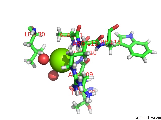
Mono view
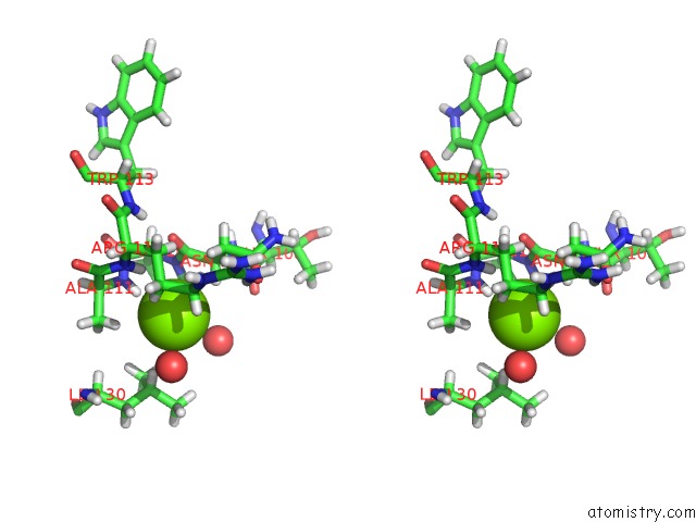
Stereo pair view
Mono view
Stereo pair view
A full contact list of Magnesium with other atoms in the Mg binding
site number 1 of Crystal Structure of Mycobacterium Tuberculosis RV1816 Transcriptional Regulator. within 5.0Å range:
|
Magnesium binding site 2 out of 2 in 5d1r
Go back to
Magnesium binding site 2 out
of 2 in the Crystal Structure of Mycobacterium Tuberculosis RV1816 Transcriptional Regulator.
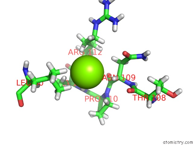
Mono view
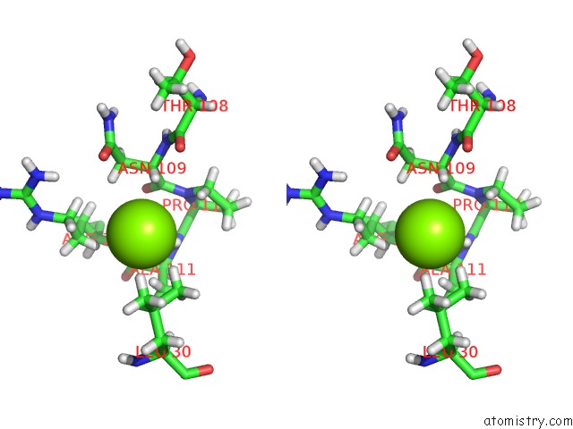
Stereo pair view
Mono view
Stereo pair view
A full contact list of Magnesium with other atoms in the Mg binding
site number 2 of Crystal Structure of Mycobacterium Tuberculosis RV1816 Transcriptional Regulator. within 5.0Å range:
|
Reference:
J.A.Delmar,
T.H.Chou,
C.C.Wright,
M.H.Licon,
J.K.Doh,
A.Radhakrishnan,
N.Kumar,
H.T.Lei,
J.R.Bolla,
K.R.Rajashankar,
C.C.Su,
G.E.Purdy,
E.W.Yu.
Structural Basis For the Regulation of the Mmpl Transporters of Mycobacterium Tuberculosis. J.Biol.Chem. V. 290 28559 2015.
ISSN: ESSN 1083-351X
PubMed: 26396194
DOI: 10.1074/JBC.M115.683797
Page generated: Sun Sep 29 02:27:49 2024
ISSN: ESSN 1083-351X
PubMed: 26396194
DOI: 10.1074/JBC.M115.683797
Last articles
Fe in 2YXOFe in 2YRS
Fe in 2YXC
Fe in 2YNM
Fe in 2YVJ
Fe in 2YP1
Fe in 2YU2
Fe in 2YU1
Fe in 2YQB
Fe in 2YOO