Magnesium »
PDB 5d2k-5dar »
5d5v »
Magnesium in PDB 5d5v: Crystal Structure of Human HSF1 with Satellite III Repeat Dna
Protein crystallography data
The structure of Crystal Structure of Human HSF1 with Satellite III Repeat Dna, PDB code: 5d5v
was solved by
T.Neudegger,
J.Verghese,
M.Hayer-Hartl,
F.U.Hartl,
A.Bracher,
with X-Ray Crystallography technique. A brief refinement statistics is given in the table below:
| Resolution Low / High (Å) | 30.00 / 2.55 |
| Space group | C 1 2 1 |
| Cell size a, b, c (Å), α, β, γ (°) | 104.813, 112.678, 39.865, 90.00, 92.56, 90.00 |
| R / Rfree (%) | 19 / 23.4 |
Magnesium Binding Sites:
The binding sites of Magnesium atom in the Crystal Structure of Human HSF1 with Satellite III Repeat Dna
(pdb code 5d5v). This binding sites where shown within
5.0 Angstroms radius around Magnesium atom.
In total 2 binding sites of Magnesium where determined in the Crystal Structure of Human HSF1 with Satellite III Repeat Dna, PDB code: 5d5v:
Jump to Magnesium binding site number: 1; 2;
In total 2 binding sites of Magnesium where determined in the Crystal Structure of Human HSF1 with Satellite III Repeat Dna, PDB code: 5d5v:
Jump to Magnesium binding site number: 1; 2;
Magnesium binding site 1 out of 2 in 5d5v
Go back to
Magnesium binding site 1 out
of 2 in the Crystal Structure of Human HSF1 with Satellite III Repeat Dna
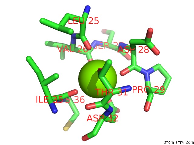
Mono view
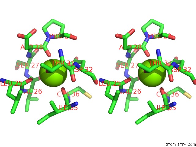
Stereo pair view
Mono view
Stereo pair view
A full contact list of Magnesium with other atoms in the Mg binding
site number 1 of Crystal Structure of Human HSF1 with Satellite III Repeat Dna within 5.0Å range:
|
Magnesium binding site 2 out of 2 in 5d5v
Go back to
Magnesium binding site 2 out
of 2 in the Crystal Structure of Human HSF1 with Satellite III Repeat Dna
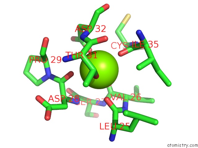
Mono view
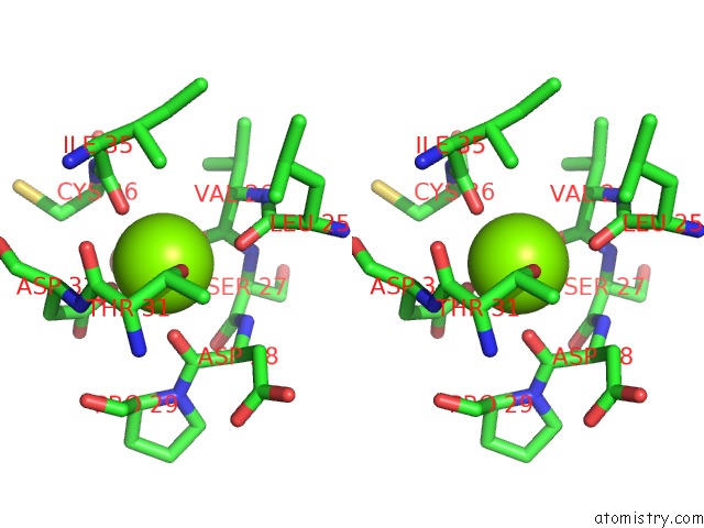
Stereo pair view
Mono view
Stereo pair view
A full contact list of Magnesium with other atoms in the Mg binding
site number 2 of Crystal Structure of Human HSF1 with Satellite III Repeat Dna within 5.0Å range:
|
Reference:
T.Neudegger,
J.Verghese,
M.Hayer-Hartl,
F.U.Hartl,
A.Bracher.
Structure of Human Heat-Shock Transcription Factor 1 in Complex with Dna. Nat.Struct.Mol.Biol. V. 23 140 2016.
ISSN: ESSN 1545-9985
PubMed: 26727489
DOI: 10.1038/NSMB.3149
Page generated: Tue Aug 12 06:46:00 2025
ISSN: ESSN 1545-9985
PubMed: 26727489
DOI: 10.1038/NSMB.3149
Last articles
Mg in 6CAQMg in 6CGD
Mg in 6CFV
Mg in 6CFU
Mg in 6CFT
Mg in 6CEY
Mg in 6CFS
Mg in 6CFR
Mg in 6CFO
Mg in 6CER