Magnesium »
PDB 5d2k-5dar »
5d6j »
Magnesium in PDB 5d6j: Crystal Structure of A Mycobacterial Protein
Protein crystallography data
The structure of Crystal Structure of A Mycobacterial Protein, PDB code: 5d6j
was solved by
W.J.Li,
L.J.Bi,
with X-Ray Crystallography technique. A brief refinement statistics is given in the table below:
| Resolution Low / High (Å) | 29.99 / 2.25 |
| Space group | P 43 21 2 |
| Cell size a, b, c (Å), α, β, γ (°) | 122.211, 122.211, 142.588, 90.00, 90.00, 90.00 |
| R / Rfree (%) | 16.9 / 20.6 |
Magnesium Binding Sites:
The binding sites of Magnesium atom in the Crystal Structure of A Mycobacterial Protein
(pdb code 5d6j). This binding sites where shown within
5.0 Angstroms radius around Magnesium atom.
In total only one binding site of Magnesium was determined in the Crystal Structure of A Mycobacterial Protein, PDB code: 5d6j:
In total only one binding site of Magnesium was determined in the Crystal Structure of A Mycobacterial Protein, PDB code: 5d6j:
Magnesium binding site 1 out of 1 in 5d6j
Go back to
Magnesium binding site 1 out
of 1 in the Crystal Structure of A Mycobacterial Protein
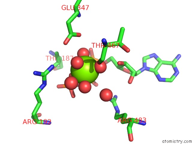
Mono view
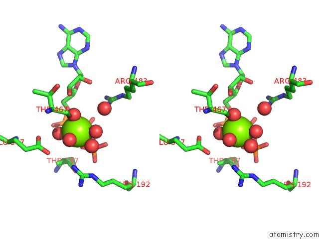
Stereo pair view
Mono view
Stereo pair view
A full contact list of Magnesium with other atoms in the Mg binding
site number 1 of Crystal Structure of A Mycobacterial Protein within 5.0Å range:
|
Reference:
W.Li,
S.Gu,
J.Fleming,
L.Bi.
Crystal Structure of FADD32, An Enzyme Essential For Mycolic Acid Biosynthesis in Mycobacteria. Sci Rep V. 5 15493 2015.
ISSN: ESSN 2045-2322
PubMed: 26628098
DOI: 10.1038/SREP15493
Page generated: Tue Aug 12 06:46:14 2025
ISSN: ESSN 2045-2322
PubMed: 26628098
DOI: 10.1038/SREP15493
Last articles
Mg in 5HKVMg in 5HR6
Mg in 5HQL
Mg in 5HQW
Mg in 5HQM
Mg in 5HQC
Mg in 5HQB
Mg in 5HQ8
Mg in 5HQA
Mg in 5HQ4