Magnesium »
PDB 5ds5-5e75 »
5dxn »
Magnesium in PDB 5dxn: Structure of Aspergillus Fumigatus Trehalose-6-Phosphate Phosphatase Crystal Form 2
Enzymatic activity of Structure of Aspergillus Fumigatus Trehalose-6-Phosphate Phosphatase Crystal Form 2
All present enzymatic activity of Structure of Aspergillus Fumigatus Trehalose-6-Phosphate Phosphatase Crystal Form 2:
2.4.1.15;
2.4.1.15;
Protein crystallography data
The structure of Structure of Aspergillus Fumigatus Trehalose-6-Phosphate Phosphatase Crystal Form 2, PDB code: 5dxn
was solved by
Y.Miao,
R.G.Brennan,
with X-Ray Crystallography technique. A brief refinement statistics is given in the table below:
| Resolution Low / High (Å) | 20.60 / 1.65 |
| Space group | P 1 21 1 |
| Cell size a, b, c (Å), α, β, γ (°) | 39.843, 42.672, 80.221, 90.00, 99.46, 90.00 |
| R / Rfree (%) | 21.9 / 25.3 |
Magnesium Binding Sites:
The binding sites of Magnesium atom in the Structure of Aspergillus Fumigatus Trehalose-6-Phosphate Phosphatase Crystal Form 2
(pdb code 5dxn). This binding sites where shown within
5.0 Angstroms radius around Magnesium atom.
In total only one binding site of Magnesium was determined in the Structure of Aspergillus Fumigatus Trehalose-6-Phosphate Phosphatase Crystal Form 2, PDB code: 5dxn:
In total only one binding site of Magnesium was determined in the Structure of Aspergillus Fumigatus Trehalose-6-Phosphate Phosphatase Crystal Form 2, PDB code: 5dxn:
Magnesium binding site 1 out of 1 in 5dxn
Go back to
Magnesium binding site 1 out
of 1 in the Structure of Aspergillus Fumigatus Trehalose-6-Phosphate Phosphatase Crystal Form 2
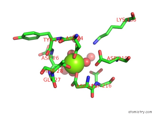
Mono view
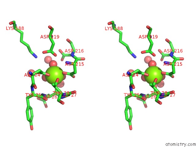
Stereo pair view
Mono view
Stereo pair view
A full contact list of Magnesium with other atoms in the Mg binding
site number 1 of Structure of Aspergillus Fumigatus Trehalose-6-Phosphate Phosphatase Crystal Form 2 within 5.0Å range:
|
Reference:
Y.Miao,
J.L.Tenor,
D.L.Toffaletti,
E.J.Washington,
J.Liu,
W.R.Shadrick,
M.A.Schumacher,
R.E.Lee,
J.R.Perfect,
R.G.Brennan.
Structures of Trehalose-6-Phosphate Phosphatase From Pathogenic Fungi Reveal the Mechanisms of Substrate Recognition and Catalysis. Proc.Natl.Acad.Sci.Usa V. 113 7148 2016.
ISSN: ESSN 1091-6490
PubMed: 27307435
DOI: 10.1073/PNAS.1601774113
Page generated: Sun Sep 29 03:26:32 2024
ISSN: ESSN 1091-6490
PubMed: 27307435
DOI: 10.1073/PNAS.1601774113
Last articles
Zn in 9J0NZn in 9J0O
Zn in 9J0P
Zn in 9FJX
Zn in 9EKB
Zn in 9C0F
Zn in 9CAH
Zn in 9CH0
Zn in 9CH3
Zn in 9CH1