Magnesium »
PDB 5ds5-5e75 »
5e41 »
Magnesium in PDB 5e41: Crystal Structure of the Large Fragment of Dna Polymerase I From Thermus Aquaticus in A Closed Ternary Complex with 5-(N-(10- Hydroxydecanoyl)-Aminopentenyl)-2'-Deoxyuridine-Triphosphate
Enzymatic activity of Crystal Structure of the Large Fragment of Dna Polymerase I From Thermus Aquaticus in A Closed Ternary Complex with 5-(N-(10- Hydroxydecanoyl)-Aminopentenyl)-2'-Deoxyuridine-Triphosphate
All present enzymatic activity of Crystal Structure of the Large Fragment of Dna Polymerase I From Thermus Aquaticus in A Closed Ternary Complex with 5-(N-(10- Hydroxydecanoyl)-Aminopentenyl)-2'-Deoxyuridine-Triphosphate:
2.7.7.7;
2.7.7.7;
Protein crystallography data
The structure of Crystal Structure of the Large Fragment of Dna Polymerase I From Thermus Aquaticus in A Closed Ternary Complex with 5-(N-(10- Hydroxydecanoyl)-Aminopentenyl)-2'-Deoxyuridine-Triphosphate, PDB code: 5e41
was solved by
A.Hottin,
K.Betz,
A.Marx,
with X-Ray Crystallography technique. A brief refinement statistics is given in the table below:
| Resolution Low / High (Å) | 46.67 / 1.80 |
| Space group | P 31 2 1 |
| Cell size a, b, c (Å), α, β, γ (°) | 107.774, 107.774, 89.614, 90.00, 90.00, 120.00 |
| R / Rfree (%) | 19.2 / 23.6 |
Magnesium Binding Sites:
The binding sites of Magnesium atom in the Crystal Structure of the Large Fragment of Dna Polymerase I From Thermus Aquaticus in A Closed Ternary Complex with 5-(N-(10- Hydroxydecanoyl)-Aminopentenyl)-2'-Deoxyuridine-Triphosphate
(pdb code 5e41). This binding sites where shown within
5.0 Angstroms radius around Magnesium atom.
In total 2 binding sites of Magnesium where determined in the Crystal Structure of the Large Fragment of Dna Polymerase I From Thermus Aquaticus in A Closed Ternary Complex with 5-(N-(10- Hydroxydecanoyl)-Aminopentenyl)-2'-Deoxyuridine-Triphosphate, PDB code: 5e41:
Jump to Magnesium binding site number: 1; 2;
Magnesium binding site 1 out of 2 in 5e41
Go back to
Magnesium binding site 1 out
of 2 in the Crystal Structure of the Large Fragment of Dna Polymerase I From Thermus Aquaticus in A Closed Ternary Complex with 5-(N-(10- Hydroxydecanoyl)-Aminopentenyl)-2'-Deoxyuridine-Triphosphate
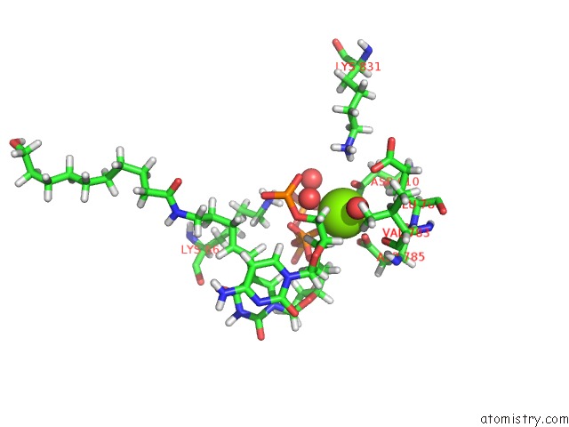
Mono view
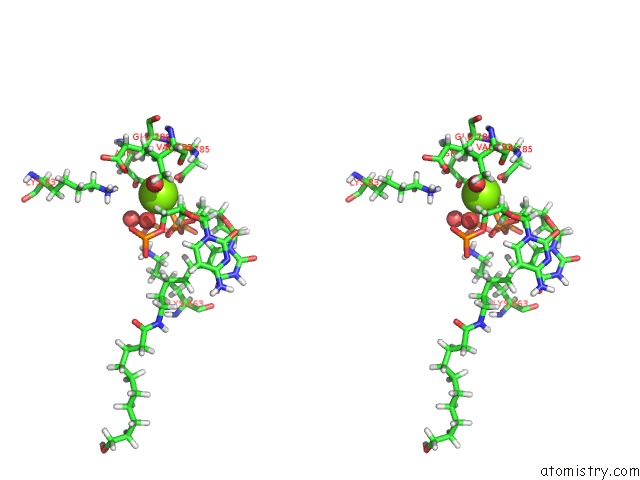
Stereo pair view
Mono view
Stereo pair view
A full contact list of Magnesium with other atoms in the Mg binding
site number 1 of Crystal Structure of the Large Fragment of Dna Polymerase I From Thermus Aquaticus in A Closed Ternary Complex with 5-(N-(10- Hydroxydecanoyl)-Aminopentenyl)-2'-Deoxyuridine-Triphosphate within 5.0Å range:
|
Magnesium binding site 2 out of 2 in 5e41
Go back to
Magnesium binding site 2 out
of 2 in the Crystal Structure of the Large Fragment of Dna Polymerase I From Thermus Aquaticus in A Closed Ternary Complex with 5-(N-(10- Hydroxydecanoyl)-Aminopentenyl)-2'-Deoxyuridine-Triphosphate
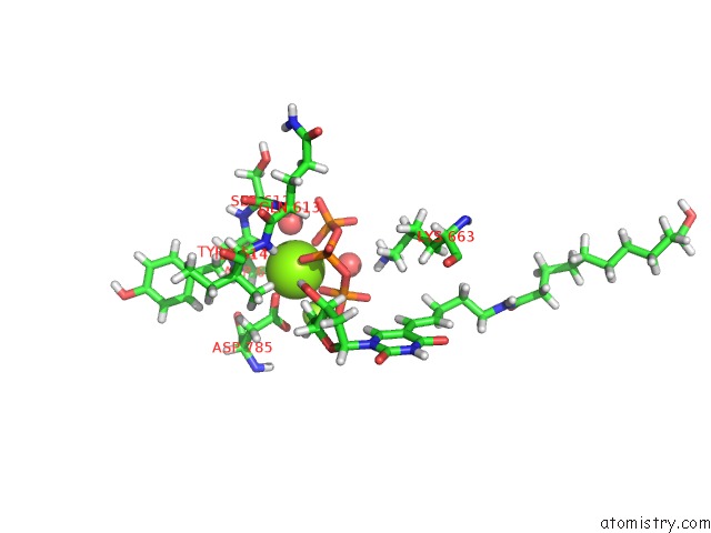
Mono view
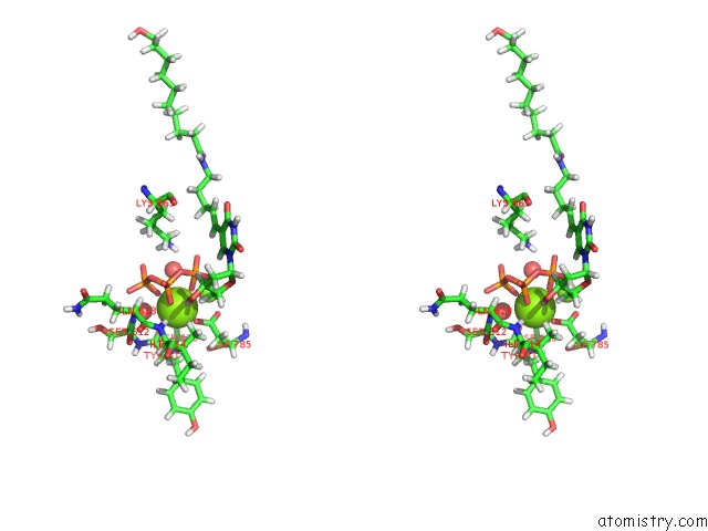
Stereo pair view
Mono view
Stereo pair view
A full contact list of Magnesium with other atoms in the Mg binding
site number 2 of Crystal Structure of the Large Fragment of Dna Polymerase I From Thermus Aquaticus in A Closed Ternary Complex with 5-(N-(10- Hydroxydecanoyl)-Aminopentenyl)-2'-Deoxyuridine-Triphosphate within 5.0Å range:
|
Reference:
A.Hottin,
K.Betz,
K.Diederichs,
A.Marx.
Structural Basis For the Klentaq Dna Polymerase Catalysed Incorporation of Alkene- Versus Alkyne-Modified Nucleotides. Chemistry V. 23 2109 2017.
ISSN: ISSN 1521-3765
PubMed: 27901305
DOI: 10.1002/CHEM.201604515
Page generated: Sun Sep 29 03:29:30 2024
ISSN: ISSN 1521-3765
PubMed: 27901305
DOI: 10.1002/CHEM.201604515
Last articles
Zn in 9J0NZn in 9J0O
Zn in 9J0P
Zn in 9FJX
Zn in 9EKB
Zn in 9C0F
Zn in 9CAH
Zn in 9CH0
Zn in 9CH3
Zn in 9CH1