Magnesium »
PDB 5e79-5eg3 »
5ect »
Magnesium in PDB 5ect: Mycobacterium Tuberculosis Dutpase G143STOP Mutant
Enzymatic activity of Mycobacterium Tuberculosis Dutpase G143STOP Mutant
All present enzymatic activity of Mycobacterium Tuberculosis Dutpase G143STOP Mutant:
3.6.1.23;
3.6.1.23;
Protein crystallography data
The structure of Mycobacterium Tuberculosis Dutpase G143STOP Mutant, PDB code: 5ect
was solved by
G.N.Nagy,
I.Leveles,
V.Harmat,
G.B.Vertessy,
with X-Ray Crystallography technique. A brief refinement statistics is given in the table below:
| Resolution Low / High (Å) | 26.04 / 1.30 |
| Space group | P 63 |
| Cell size a, b, c (Å), α, β, γ (°) | 54.780, 54.780, 84.079, 90.00, 90.00, 120.00 |
| R / Rfree (%) | 12.3 / 15.7 |
Magnesium Binding Sites:
The binding sites of Magnesium atom in the Mycobacterium Tuberculosis Dutpase G143STOP Mutant
(pdb code 5ect). This binding sites where shown within
5.0 Angstroms radius around Magnesium atom.
In total only one binding site of Magnesium was determined in the Mycobacterium Tuberculosis Dutpase G143STOP Mutant, PDB code: 5ect:
In total only one binding site of Magnesium was determined in the Mycobacterium Tuberculosis Dutpase G143STOP Mutant, PDB code: 5ect:
Magnesium binding site 1 out of 1 in 5ect
Go back to
Magnesium binding site 1 out
of 1 in the Mycobacterium Tuberculosis Dutpase G143STOP Mutant
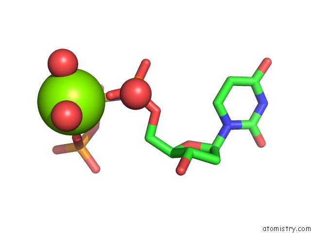
Mono view
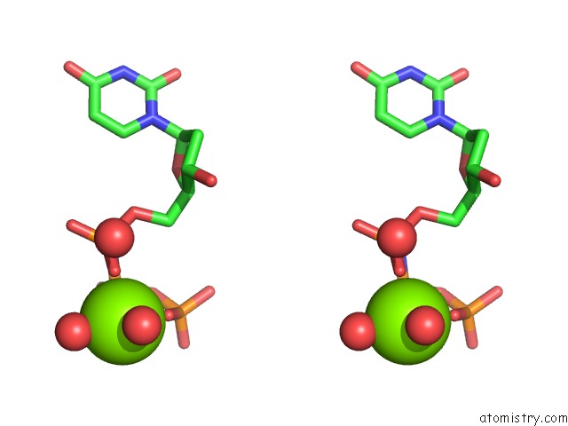
Stereo pair view
Mono view
Stereo pair view
A full contact list of Magnesium with other atoms in the Mg binding
site number 1 of Mycobacterium Tuberculosis Dutpase G143STOP Mutant within 5.0Å range:
|
Reference:
G.N.Nagy,
R.Suardiaz,
A.Lopata,
O.Ozohanics,
K.Vekey,
B.R.Brooks,
I.Leveles,
J.Toth,
B.G.Vertessy,
E.Rosta.
Structural Characterization of Arginine Fingers: Identification of An Arginine Finger For the Pyrophosphatase Dutpases. J. Am. Chem. Soc. V. 138 15035 2016.
ISSN: ESSN 1520-5126
PubMed: 27740761
DOI: 10.1021/JACS.6B09012
Page generated: Sun Sep 29 03:37:04 2024
ISSN: ESSN 1520-5126
PubMed: 27740761
DOI: 10.1021/JACS.6B09012
Last articles
Zn in 9J0NZn in 9J0O
Zn in 9J0P
Zn in 9FJX
Zn in 9EKB
Zn in 9C0F
Zn in 9CAH
Zn in 9CH0
Zn in 9CH3
Zn in 9CH1