Magnesium »
PDB 5e79-5eg3 »
5edk »
Magnesium in PDB 5edk: Crystal Structure of Prothrombin Deletion Mutant Residues 146-167 ( Form II ).
Enzymatic activity of Crystal Structure of Prothrombin Deletion Mutant Residues 146-167 ( Form II ).
All present enzymatic activity of Crystal Structure of Prothrombin Deletion Mutant Residues 146-167 ( Form II ).:
3.4.21.5;
3.4.21.5;
Protein crystallography data
The structure of Crystal Structure of Prothrombin Deletion Mutant Residues 146-167 ( Form II )., PDB code: 5edk
was solved by
N.Pozzi,
Z.Chen,
E.Di Cera,
with X-Ray Crystallography technique. A brief refinement statistics is given in the table below:
| Resolution Low / High (Å) | 86.61 / 3.21 |
| Space group | P 41 21 2 |
| Cell size a, b, c (Å), α, β, γ (°) | 84.192, 84.192, 346.427, 90.00, 90.00, 90.00 |
| R / Rfree (%) | 29.1 / 32.3 |
Magnesium Binding Sites:
The binding sites of Magnesium atom in the Crystal Structure of Prothrombin Deletion Mutant Residues 146-167 ( Form II ).
(pdb code 5edk). This binding sites where shown within
5.0 Angstroms radius around Magnesium atom.
In total 4 binding sites of Magnesium where determined in the Crystal Structure of Prothrombin Deletion Mutant Residues 146-167 ( Form II )., PDB code: 5edk:
Jump to Magnesium binding site number: 1; 2; 3; 4;
In total 4 binding sites of Magnesium where determined in the Crystal Structure of Prothrombin Deletion Mutant Residues 146-167 ( Form II )., PDB code: 5edk:
Jump to Magnesium binding site number: 1; 2; 3; 4;
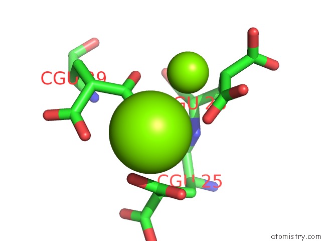
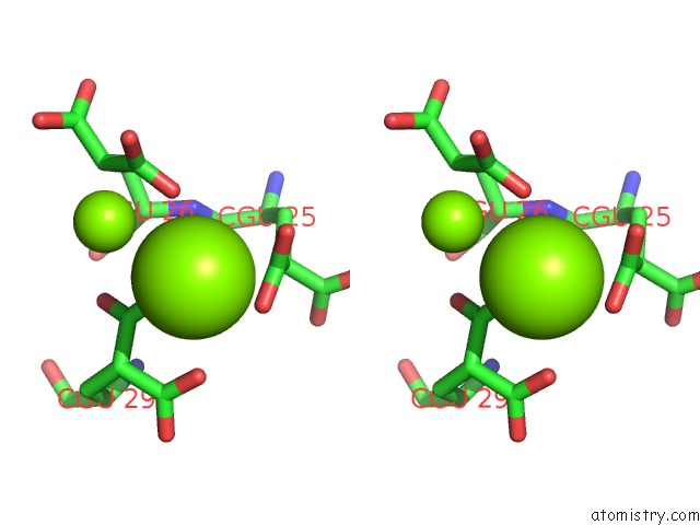
Magnesium binding site 1 out of 4 in 5edk
Go back to
Magnesium binding site 1 out
of 4 in the Crystal Structure of Prothrombin Deletion Mutant Residues 146-167 ( Form II ).
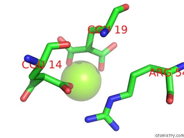
Mono view
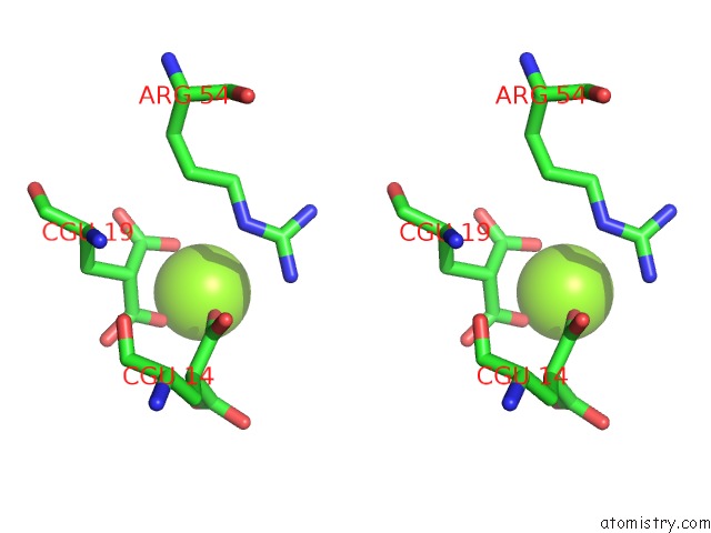
Stereo pair view
Mono view
Stereo pair view
A full contact list of Magnesium with other atoms in the Mg binding
site number 1 of Crystal Structure of Prothrombin Deletion Mutant Residues 146-167 ( Form II ). within 5.0Å range:
|
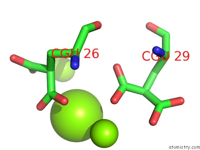
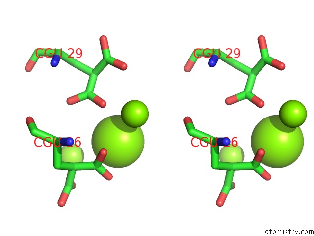
Magnesium binding site 2 out of 4 in 5edk
Go back to
Magnesium binding site 2 out
of 4 in the Crystal Structure of Prothrombin Deletion Mutant Residues 146-167 ( Form II ).
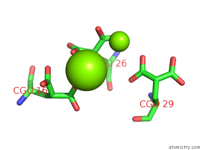
Mono view
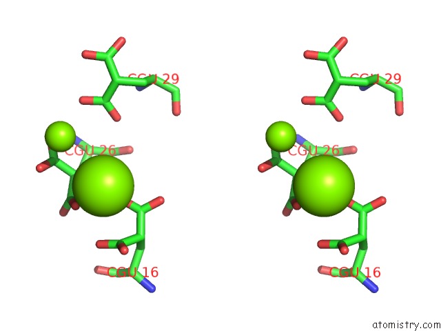
Stereo pair view
Mono view
Stereo pair view
A full contact list of Magnesium with other atoms in the Mg binding
site number 2 of Crystal Structure of Prothrombin Deletion Mutant Residues 146-167 ( Form II ). within 5.0Å range:
|
Magnesium binding site 3 out of 4 in 5edk
Go back to
Magnesium binding site 3 out
of 4 in the Crystal Structure of Prothrombin Deletion Mutant Residues 146-167 ( Form II ).
Mono view
Stereo pair view
Mono view
Stereo pair view
A full contact list of Magnesium with other atoms in the Mg binding
site number 3 of Crystal Structure of Prothrombin Deletion Mutant Residues 146-167 ( Form II ). within 5.0Å range:
|
Magnesium binding site 4 out of 4 in 5edk
Go back to
Magnesium binding site 4 out
of 4 in the Crystal Structure of Prothrombin Deletion Mutant Residues 146-167 ( Form II ).
Mono view
Stereo pair view
Mono view
Stereo pair view
A full contact list of Magnesium with other atoms in the Mg binding
site number 4 of Crystal Structure of Prothrombin Deletion Mutant Residues 146-167 ( Form II ). within 5.0Å range:
|
Reference:
N.Pozzi,
Z.Chen,
E.Di Cera.
How the Linker Connecting the Two Kringles Influences Activation and Conformational Plasticity of Prothrombin. J.Biol.Chem. V. 291 6071 2016.
ISSN: ESSN 1083-351X
PubMed: 26763231
DOI: 10.1074/JBC.M115.700401
Page generated: Sun Sep 29 03:38:12 2024
ISSN: ESSN 1083-351X
PubMed: 26763231
DOI: 10.1074/JBC.M115.700401
Last articles
Zn in 9J0NZn in 9J0O
Zn in 9J0P
Zn in 9FJX
Zn in 9EKB
Zn in 9C0F
Zn in 9CAH
Zn in 9CH0
Zn in 9CH3
Zn in 9CH1