Magnesium »
PDB 5ets-5f7u »
5ex2 »
Magnesium in PDB 5ex2: Crystal Structure of Cyclophilin AQUACYP293 From Hirschia Baltica
Enzymatic activity of Crystal Structure of Cyclophilin AQUACYP293 From Hirschia Baltica
All present enzymatic activity of Crystal Structure of Cyclophilin AQUACYP293 From Hirschia Baltica:
5.2.1.8;
5.2.1.8;
Protein crystallography data
The structure of Crystal Structure of Cyclophilin AQUACYP293 From Hirschia Baltica, PDB code: 5ex2
was solved by
R.P.Jakob,
T.Maier,
with X-Ray Crystallography technique. A brief refinement statistics is given in the table below:
| Resolution Low / High (Å) | 47.87 / 1.29 |
| Space group | P 1 21 1 |
| Cell size a, b, c (Å), α, β, γ (°) | 47.940, 72.730, 73.930, 90.00, 93.00, 90.00 |
| R / Rfree (%) | 16.9 / 19.5 |
Other elements in 5ex2:
The structure of Crystal Structure of Cyclophilin AQUACYP293 From Hirschia Baltica also contains other interesting chemical elements:
| Calcium | (Ca) | 1 atom |
| Chlorine | (Cl) | 1 atom |
Magnesium Binding Sites:
The binding sites of Magnesium atom in the Crystal Structure of Cyclophilin AQUACYP293 From Hirschia Baltica
(pdb code 5ex2). This binding sites where shown within
5.0 Angstroms radius around Magnesium atom.
In total 2 binding sites of Magnesium where determined in the Crystal Structure of Cyclophilin AQUACYP293 From Hirschia Baltica, PDB code: 5ex2:
Jump to Magnesium binding site number: 1; 2;
In total 2 binding sites of Magnesium where determined in the Crystal Structure of Cyclophilin AQUACYP293 From Hirschia Baltica, PDB code: 5ex2:
Jump to Magnesium binding site number: 1; 2;
Magnesium binding site 1 out of 2 in 5ex2
Go back to
Magnesium binding site 1 out
of 2 in the Crystal Structure of Cyclophilin AQUACYP293 From Hirschia Baltica
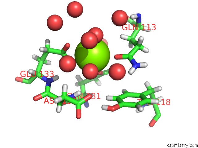
Mono view
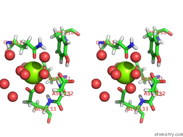
Stereo pair view
Mono view
Stereo pair view
A full contact list of Magnesium with other atoms in the Mg binding
site number 1 of Crystal Structure of Cyclophilin AQUACYP293 From Hirschia Baltica within 5.0Å range:
|
Magnesium binding site 2 out of 2 in 5ex2
Go back to
Magnesium binding site 2 out
of 2 in the Crystal Structure of Cyclophilin AQUACYP293 From Hirschia Baltica
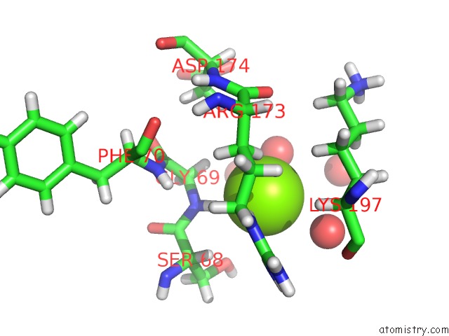
Mono view
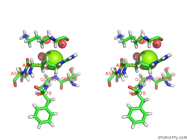
Stereo pair view
Mono view
Stereo pair view
A full contact list of Magnesium with other atoms in the Mg binding
site number 2 of Crystal Structure of Cyclophilin AQUACYP293 From Hirschia Baltica within 5.0Å range:
|
Reference:
R.P.Jakob,
P.A.Schmidpeter,
J.R.Koch,
F.X.Schmid,
T.Maier.
Structural and Functional Characterization of A Novel Family of Cyclophilins, the Aquacyps. Plos One V. 11 57070 2016.
ISSN: ESSN 1932-6203
PubMed: 27276069
DOI: 10.1371/JOURNAL.PONE.0157070
Page generated: Tue Aug 12 07:54:36 2025
ISSN: ESSN 1932-6203
PubMed: 27276069
DOI: 10.1371/JOURNAL.PONE.0157070
Last articles
Mg in 6DW3Mg in 6DUQ
Mg in 6DVK
Mg in 6DUK
Mg in 6DV9
Mg in 6DUS
Mg in 6DUH
Mg in 6DUG
Mg in 6DUF
Mg in 6DUE