Magnesium »
PDB 5etr-5f6c »
5f48 »
Magnesium in PDB 5f48: Crystal Structure of An Aminoglycoside Acetyltransferase Meta-AAC0020 From An Uncultured Soil Metagenomic Sample in Complex with Coenzyme A
Protein crystallography data
The structure of Crystal Structure of An Aminoglycoside Acetyltransferase Meta-AAC0020 From An Uncultured Soil Metagenomic Sample in Complex with Coenzyme A, PDB code: 5f48
was solved by
Z.Xu,
T.Skarina,
P.J.Stogios,
V.Yim,
A.Savchenko,
W.F.Anderson,
Center Forstructural Genomics Of Infectious Diseases (Csgid),
with X-Ray Crystallography technique. A brief refinement statistics is given in the table below:
| Resolution Low / High (Å) | 23.62 / 1.95 |
| Space group | P 1 21 1 |
| Cell size a, b, c (Å), α, β, γ (°) | 46.133, 80.187, 53.926, 90.00, 91.97, 90.00 |
| R / Rfree (%) | 19 / 23.5 |
Other elements in 5f48:
The structure of Crystal Structure of An Aminoglycoside Acetyltransferase Meta-AAC0020 From An Uncultured Soil Metagenomic Sample in Complex with Coenzyme A also contains other interesting chemical elements:
| Chlorine | (Cl) | 1 atom |
Magnesium Binding Sites:
The binding sites of Magnesium atom in the Crystal Structure of An Aminoglycoside Acetyltransferase Meta-AAC0020 From An Uncultured Soil Metagenomic Sample in Complex with Coenzyme A
(pdb code 5f48). This binding sites where shown within
5.0 Angstroms radius around Magnesium atom.
In total 3 binding sites of Magnesium where determined in the Crystal Structure of An Aminoglycoside Acetyltransferase Meta-AAC0020 From An Uncultured Soil Metagenomic Sample in Complex with Coenzyme A, PDB code: 5f48:
Jump to Magnesium binding site number: 1; 2; 3;
In total 3 binding sites of Magnesium where determined in the Crystal Structure of An Aminoglycoside Acetyltransferase Meta-AAC0020 From An Uncultured Soil Metagenomic Sample in Complex with Coenzyme A, PDB code: 5f48:
Jump to Magnesium binding site number: 1; 2; 3;
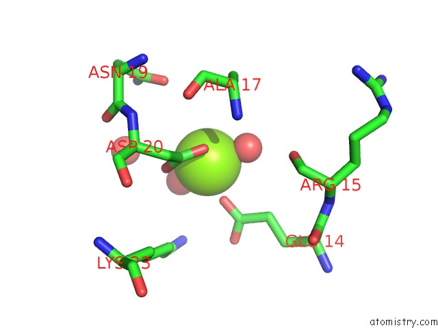
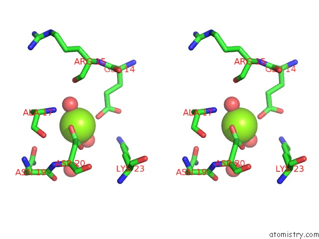
Magnesium binding site 1 out of 3 in 5f48
Go back to
Magnesium binding site 1 out
of 3 in the Crystal Structure of An Aminoglycoside Acetyltransferase Meta-AAC0020 From An Uncultured Soil Metagenomic Sample in Complex with Coenzyme A
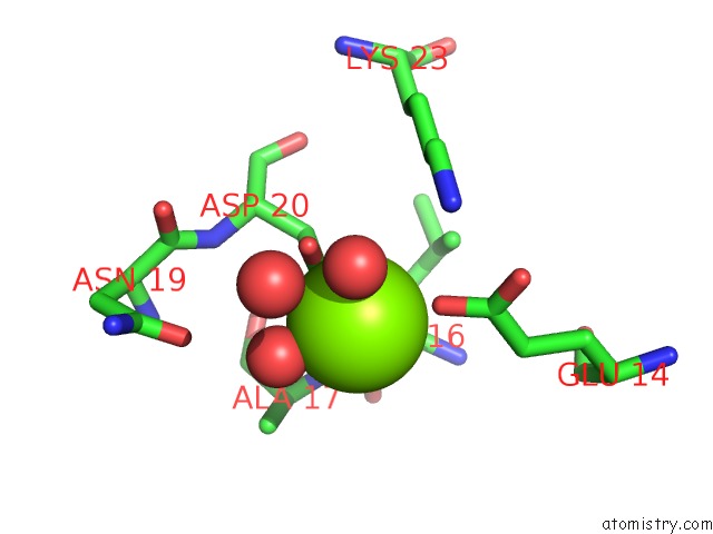
Mono view
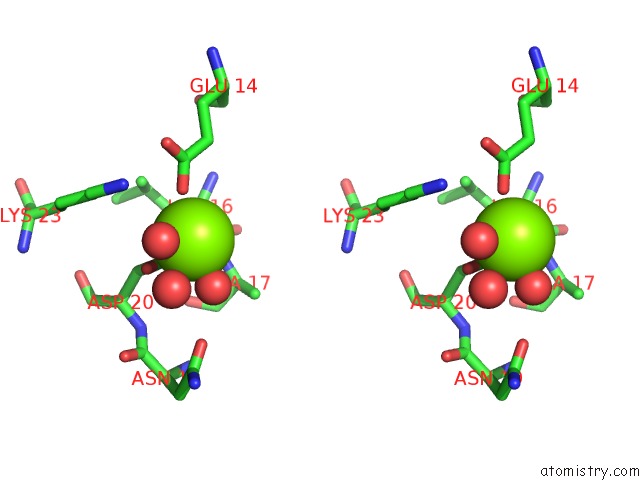
Stereo pair view
Mono view
Stereo pair view
A full contact list of Magnesium with other atoms in the Mg binding
site number 1 of Crystal Structure of An Aminoglycoside Acetyltransferase Meta-AAC0020 From An Uncultured Soil Metagenomic Sample in Complex with Coenzyme A within 5.0Å range:
|
Magnesium binding site 2 out of 3 in 5f48
Go back to
Magnesium binding site 2 out
of 3 in the Crystal Structure of An Aminoglycoside Acetyltransferase Meta-AAC0020 From An Uncultured Soil Metagenomic Sample in Complex with Coenzyme A
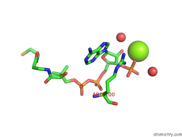
Mono view
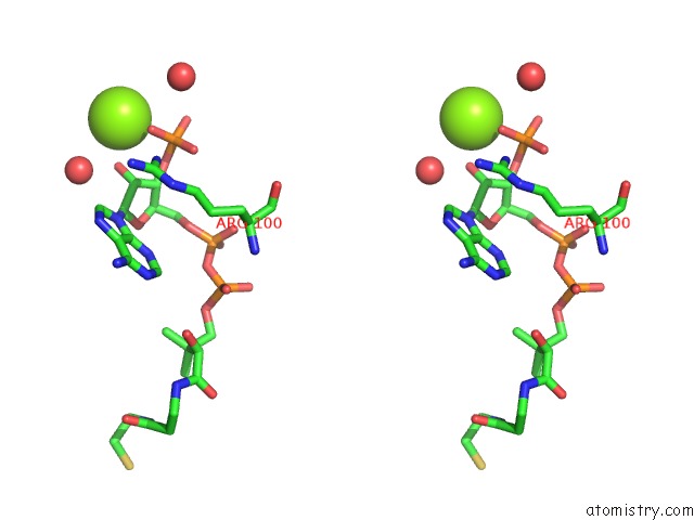
Stereo pair view
Mono view
Stereo pair view
A full contact list of Magnesium with other atoms in the Mg binding
site number 2 of Crystal Structure of An Aminoglycoside Acetyltransferase Meta-AAC0020 From An Uncultured Soil Metagenomic Sample in Complex with Coenzyme A within 5.0Å range:
|
Magnesium binding site 3 out of 3 in 5f48
Go back to
Magnesium binding site 3 out
of 3 in the Crystal Structure of An Aminoglycoside Acetyltransferase Meta-AAC0020 From An Uncultured Soil Metagenomic Sample in Complex with Coenzyme A
Mono view
Stereo pair view
Mono view
Stereo pair view
A full contact list of Magnesium with other atoms in the Mg binding
site number 3 of Crystal Structure of An Aminoglycoside Acetyltransferase Meta-AAC0020 From An Uncultured Soil Metagenomic Sample in Complex with Coenzyme A within 5.0Å range:
|
Reference:
Z.Xu,
P.J.Stogios,
A.T.Quaile,
K.J.Forsberg,
S.Patel,
T.Skarina,
S.Houliston,
C.Arrowsmith,
G.Dantas,
A.Savchenko.
Structural and Functional Survey of Environmental Aminoglycoside Acetyltransferases Reveals Functionality of Resistance Enzymes. Acs Infect Dis V. 3 653 2017.
ISSN: ESSN 2373-8227
PubMed: 28756664
DOI: 10.1021/ACSINFECDIS.7B00068
Page generated: Sun Sep 29 04:03:18 2024
ISSN: ESSN 2373-8227
PubMed: 28756664
DOI: 10.1021/ACSINFECDIS.7B00068
Last articles
Zn in 9J0NZn in 9J0O
Zn in 9J0P
Zn in 9FJX
Zn in 9EKB
Zn in 9C0F
Zn in 9CAH
Zn in 9CH0
Zn in 9CH3
Zn in 9CH1