Magnesium »
PDB 5f8h-5fjo »
5fbh »
Magnesium in PDB 5fbh: Crystal Structure of the Extracellular Domain of Human Calcium Sensing Receptor with Bound GD3+
Protein crystallography data
The structure of Crystal Structure of the Extracellular Domain of Human Calcium Sensing Receptor with Bound GD3+, PDB code: 5fbh
was solved by
T.Zhang,
C.Zhang,
C.L.Miller,
J.Zou,
K.W.Moremen,
E.M.Brown,
J.J.Yang,
J.Hu,
with X-Ray Crystallography technique. A brief refinement statistics is given in the table below:
| Resolution Low / High (Å) | 46.08 / 2.70 |
| Space group | C 1 2 1 |
| Cell size a, b, c (Å), α, β, γ (°) | 172.107, 83.106, 94.470, 90.00, 105.15, 90.00 |
| R / Rfree (%) | 18.2 / 23.6 |
Other elements in 5fbh:
The structure of Crystal Structure of the Extracellular Domain of Human Calcium Sensing Receptor with Bound GD3+ also contains other interesting chemical elements:
| Gadolinium | (Gd) | 2 atoms |
| Chlorine | (Cl) | 2 atoms |
Magnesium Binding Sites:
The binding sites of Magnesium atom in the Crystal Structure of the Extracellular Domain of Human Calcium Sensing Receptor with Bound GD3+
(pdb code 5fbh). This binding sites where shown within
5.0 Angstroms radius around Magnesium atom.
In total 3 binding sites of Magnesium where determined in the Crystal Structure of the Extracellular Domain of Human Calcium Sensing Receptor with Bound GD3+, PDB code: 5fbh:
Jump to Magnesium binding site number: 1; 2; 3;
In total 3 binding sites of Magnesium where determined in the Crystal Structure of the Extracellular Domain of Human Calcium Sensing Receptor with Bound GD3+, PDB code: 5fbh:
Jump to Magnesium binding site number: 1; 2; 3;
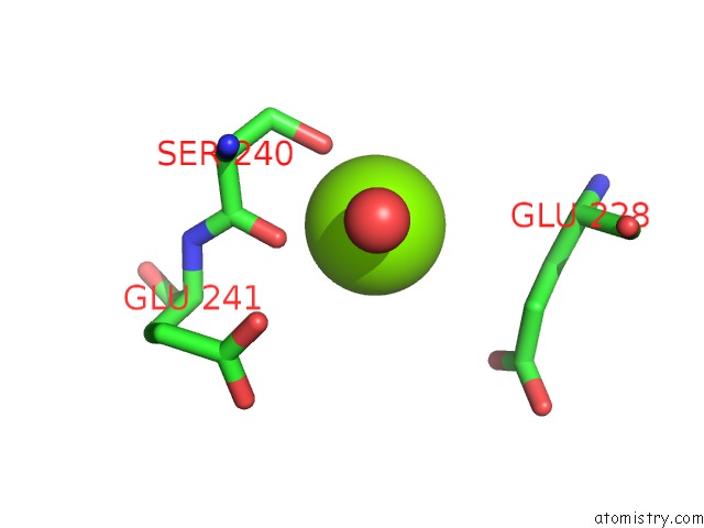
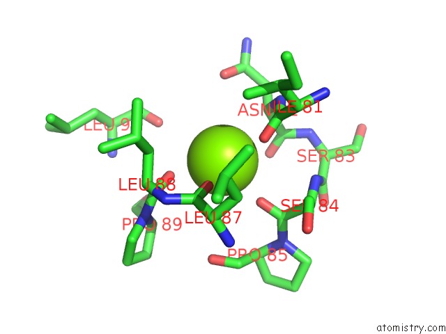
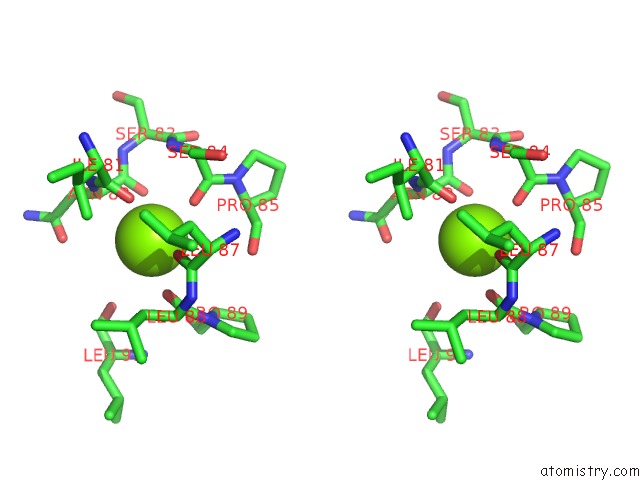
Magnesium binding site 1 out of 3 in 5fbh
Go back to
Magnesium binding site 1 out
of 3 in the Crystal Structure of the Extracellular Domain of Human Calcium Sensing Receptor with Bound GD3+
Mono view
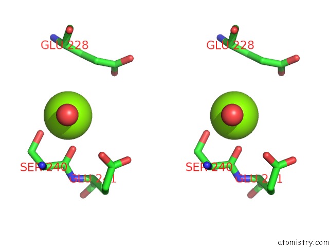
Stereo pair view
Mono view
Stereo pair view
A full contact list of Magnesium with other atoms in the Mg binding
site number 1 of Crystal Structure of the Extracellular Domain of Human Calcium Sensing Receptor with Bound GD3+ within 5.0Å range:
|
Magnesium binding site 2 out of 3 in 5fbh
Go back to
Magnesium binding site 2 out
of 3 in the Crystal Structure of the Extracellular Domain of Human Calcium Sensing Receptor with Bound GD3+
Mono view
Stereo pair view
Mono view
Stereo pair view
A full contact list of Magnesium with other atoms in the Mg binding
site number 2 of Crystal Structure of the Extracellular Domain of Human Calcium Sensing Receptor with Bound GD3+ within 5.0Å range:
|
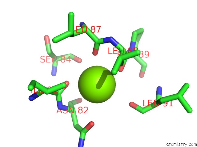
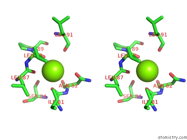
Magnesium binding site 3 out of 3 in 5fbh
Go back to
Magnesium binding site 3 out
of 3 in the Crystal Structure of the Extracellular Domain of Human Calcium Sensing Receptor with Bound GD3+
Mono view
Stereo pair view
Mono view
Stereo pair view
A full contact list of Magnesium with other atoms in the Mg binding
site number 3 of Crystal Structure of the Extracellular Domain of Human Calcium Sensing Receptor with Bound GD3+ within 5.0Å range:
|
Reference:
C.Zhang,
T.Zhang,
J.Zou,
C.L.Miller,
R.Gorkhali,
J.Y.Yang,
A.Schilmiller,
S.Wang,
K.Huang,
E.M.Brown,
K.W.Moremen,
J.Hu,
J.J.Yang.
Structural Basis For Regulation of Human Calcium-Sensing Receptor By Magnesium Ions and An Unexpected Tryptophan Derivative Co-Agonist. Sci Adv V. 2 00241 2016.
ISSN: ESSN 2375-2548
PubMed: 27386547
DOI: 10.1126/SCIADV.1600241
Page generated: Sun Sep 29 04:08:56 2024
ISSN: ESSN 2375-2548
PubMed: 27386547
DOI: 10.1126/SCIADV.1600241
Last articles
Fe in 2YXOFe in 2YRS
Fe in 2YXC
Fe in 2YNM
Fe in 2YVJ
Fe in 2YP1
Fe in 2YU2
Fe in 2YU1
Fe in 2YQB
Fe in 2YOO