Magnesium »
PDB 5fjo-5fuk »
5flg »
Magnesium in PDB 5flg: Crystal Structure of the 6-Carboxyhexanoate-Coa Ligase (Biow)From Bacillus Subtilis in Complex with Amppnp
Enzymatic activity of Crystal Structure of the 6-Carboxyhexanoate-Coa Ligase (Biow)From Bacillus Subtilis in Complex with Amppnp
All present enzymatic activity of Crystal Structure of the 6-Carboxyhexanoate-Coa Ligase (Biow)From Bacillus Subtilis in Complex with Amppnp:
6.2.1.14;
6.2.1.14;
Protein crystallography data
The structure of Crystal Structure of the 6-Carboxyhexanoate-Coa Ligase (Biow)From Bacillus Subtilis in Complex with Amppnp, PDB code: 5flg
was solved by
L.Moynie,
M.Wang,
D.J.Campopiano,
J.H.Naismith,
with X-Ray Crystallography technique. A brief refinement statistics is given in the table below:
| Resolution Low / High (Å) | 41.53 / 2.04 |
| Space group | P 21 21 21 |
| Cell size a, b, c (Å), α, β, γ (°) | 49.590, 77.900, 166.000, 90.00, 90.00, 90.00 |
| R / Rfree (%) | 20.2 / 23.2 |
Magnesium Binding Sites:
The binding sites of Magnesium atom in the Crystal Structure of the 6-Carboxyhexanoate-Coa Ligase (Biow)From Bacillus Subtilis in Complex with Amppnp
(pdb code 5flg). This binding sites where shown within
5.0 Angstroms radius around Magnesium atom.
In total 4 binding sites of Magnesium where determined in the Crystal Structure of the 6-Carboxyhexanoate-Coa Ligase (Biow)From Bacillus Subtilis in Complex with Amppnp, PDB code: 5flg:
Jump to Magnesium binding site number: 1; 2; 3; 4;
In total 4 binding sites of Magnesium where determined in the Crystal Structure of the 6-Carboxyhexanoate-Coa Ligase (Biow)From Bacillus Subtilis in Complex with Amppnp, PDB code: 5flg:
Jump to Magnesium binding site number: 1; 2; 3; 4;
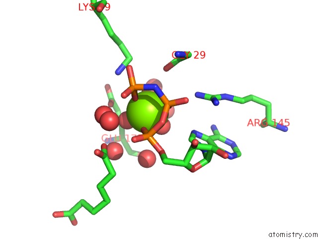
Magnesium binding site 1 out of 4 in 5flg
Go back to
Magnesium binding site 1 out
of 4 in the Crystal Structure of the 6-Carboxyhexanoate-Coa Ligase (Biow)From Bacillus Subtilis in Complex with Amppnp
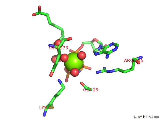
Mono view
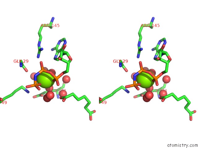
Stereo pair view
Mono view
Stereo pair view
A full contact list of Magnesium with other atoms in the Mg binding
site number 1 of Crystal Structure of the 6-Carboxyhexanoate-Coa Ligase (Biow)From Bacillus Subtilis in Complex with Amppnp within 5.0Å range:
|
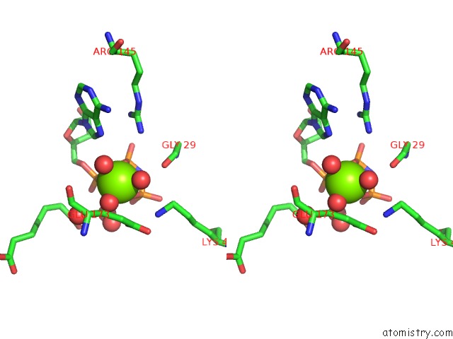
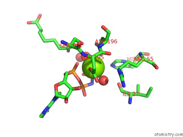
Magnesium binding site 2 out of 4 in 5flg
Go back to
Magnesium binding site 2 out
of 4 in the Crystal Structure of the 6-Carboxyhexanoate-Coa Ligase (Biow)From Bacillus Subtilis in Complex with Amppnp
Mono view
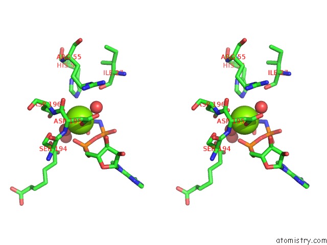
Stereo pair view
Mono view
Stereo pair view
A full contact list of Magnesium with other atoms in the Mg binding
site number 2 of Crystal Structure of the 6-Carboxyhexanoate-Coa Ligase (Biow)From Bacillus Subtilis in Complex with Amppnp within 5.0Å range:
|
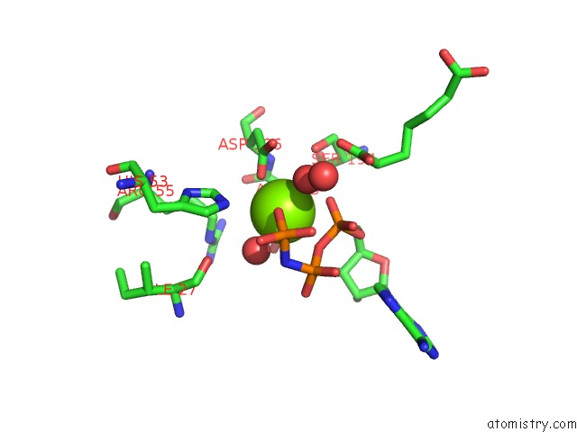
Magnesium binding site 3 out of 4 in 5flg
Go back to
Magnesium binding site 3 out
of 4 in the Crystal Structure of the 6-Carboxyhexanoate-Coa Ligase (Biow)From Bacillus Subtilis in Complex with Amppnp
Mono view
Stereo pair view
Mono view
Stereo pair view
A full contact list of Magnesium with other atoms in the Mg binding
site number 3 of Crystal Structure of the 6-Carboxyhexanoate-Coa Ligase (Biow)From Bacillus Subtilis in Complex with Amppnp within 5.0Å range:
|
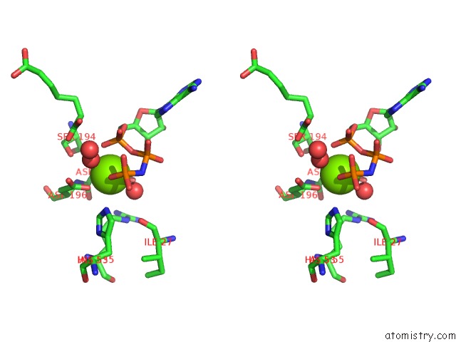
Magnesium binding site 4 out of 4 in 5flg
Go back to
Magnesium binding site 4 out
of 4 in the Crystal Structure of the 6-Carboxyhexanoate-Coa Ligase (Biow)From Bacillus Subtilis in Complex with Amppnp
Mono view
Stereo pair view
Mono view
Stereo pair view
A full contact list of Magnesium with other atoms in the Mg binding
site number 4 of Crystal Structure of the 6-Carboxyhexanoate-Coa Ligase (Biow)From Bacillus Subtilis in Complex with Amppnp within 5.0Å range:
|
Reference:
M.Wang,
L.Moynie,
P.J.Harrison,
V.Kelly,
A.Piper,
J.H.Naismith,
D.J.Campopiano.
Using the Pimeloyl-Coa Synthetase Adenylation Fold to Synthesize Fatty Acid Thioesters. Nat. Chem. Biol. V. 13 660 2017.
ISSN: ESSN 1552-4469
PubMed: 28414710
DOI: 10.1038/NCHEMBIO.2361
Page generated: Sun Sep 29 04:19:42 2024
ISSN: ESSN 1552-4469
PubMed: 28414710
DOI: 10.1038/NCHEMBIO.2361
Last articles
Zn in 9J0NZn in 9J0O
Zn in 9J0P
Zn in 9FJX
Zn in 9EKB
Zn in 9C0F
Zn in 9CAH
Zn in 9CH0
Zn in 9CH3
Zn in 9CH1