Magnesium »
PDB 5fjp-5fux »
5fml »
Magnesium in PDB 5fml: Crystal Structure of the Endonuclease From the Pa Subunit of Influenza B Virus Bound to the PB2 Subunit Nls Peptide
Protein crystallography data
The structure of Crystal Structure of the Endonuclease From the Pa Subunit of Influenza B Virus Bound to the PB2 Subunit Nls Peptide, PDB code: 5fml
was solved by
D.Guilligay,
S.Gaudon,
S.Cusack,
with X-Ray Crystallography technique. A brief refinement statistics is given in the table below:
| Resolution Low / High (Å) | 40.42 / 1.70 |
| Space group | P 1 21 1 |
| Cell size a, b, c (Å), α, β, γ (°) | 35.380, 80.850, 37.910, 90.00, 97.15, 90.00 |
| R / Rfree (%) | 22.435 / 27.556 |
Magnesium Binding Sites:
The binding sites of Magnesium atom in the Crystal Structure of the Endonuclease From the Pa Subunit of Influenza B Virus Bound to the PB2 Subunit Nls Peptide
(pdb code 5fml). This binding sites where shown within
5.0 Angstroms radius around Magnesium atom.
In total 2 binding sites of Magnesium where determined in the Crystal Structure of the Endonuclease From the Pa Subunit of Influenza B Virus Bound to the PB2 Subunit Nls Peptide, PDB code: 5fml:
Jump to Magnesium binding site number: 1; 2;
In total 2 binding sites of Magnesium where determined in the Crystal Structure of the Endonuclease From the Pa Subunit of Influenza B Virus Bound to the PB2 Subunit Nls Peptide, PDB code: 5fml:
Jump to Magnesium binding site number: 1; 2;
Magnesium binding site 1 out of 2 in 5fml
Go back to
Magnesium binding site 1 out
of 2 in the Crystal Structure of the Endonuclease From the Pa Subunit of Influenza B Virus Bound to the PB2 Subunit Nls Peptide
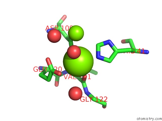
Mono view
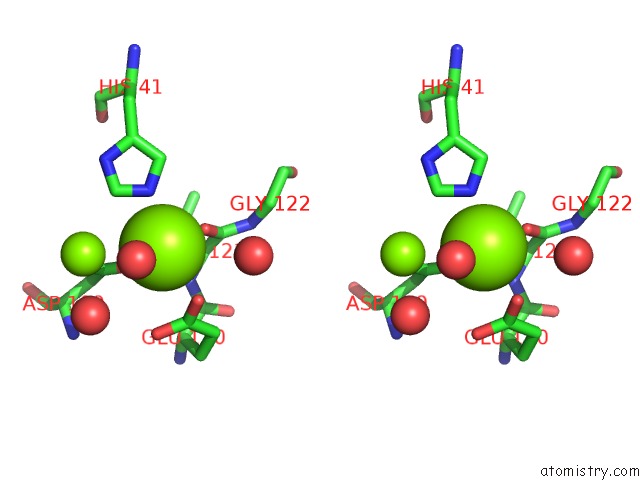
Stereo pair view
Mono view
Stereo pair view
A full contact list of Magnesium with other atoms in the Mg binding
site number 1 of Crystal Structure of the Endonuclease From the Pa Subunit of Influenza B Virus Bound to the PB2 Subunit Nls Peptide within 5.0Å range:
|
Magnesium binding site 2 out of 2 in 5fml
Go back to
Magnesium binding site 2 out
of 2 in the Crystal Structure of the Endonuclease From the Pa Subunit of Influenza B Virus Bound to the PB2 Subunit Nls Peptide
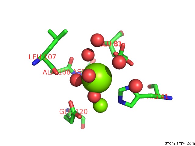
Mono view
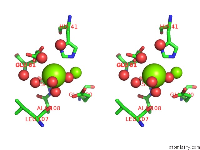
Stereo pair view
Mono view
Stereo pair view
A full contact list of Magnesium with other atoms in the Mg binding
site number 2 of Crystal Structure of the Endonuclease From the Pa Subunit of Influenza B Virus Bound to the PB2 Subunit Nls Peptide within 5.0Å range:
|
Reference:
E.Thierry,
D.Guilligay,
J.Kosinski,
T.Bock,
S.Gaudon,
A.Round,
A.Pflug,
N.Hengrung,
K.El Omari,
F.Baudin,
D.J.Hart,
M.Beck,
S.Cusack.
Influenza Polymerase Can Adopt An Alternative Configuration Involving A Radical Repacking of PB2 Domains. Mol.Cell V. 61 125 2016.
ISSN: ISSN 1097-2765
PubMed: 26711008
DOI: 10.1016/J.MOLCEL.2015.11.016
Page generated: Sun Sep 29 04:21:38 2024
ISSN: ISSN 1097-2765
PubMed: 26711008
DOI: 10.1016/J.MOLCEL.2015.11.016
Last articles
Fe in 2YXOFe in 2YRS
Fe in 2YXC
Fe in 2YNM
Fe in 2YVJ
Fe in 2YP1
Fe in 2YU2
Fe in 2YU1
Fe in 2YQB
Fe in 2YOO