Magnesium »
PDB 5fv7-5gg8 »
5fw3 »
Magnesium in PDB 5fw3: Crystal Structure of SPCAS9 Variant Vrer Bound to Sgrna and Tgcg Pam Target Dna
Protein crystallography data
The structure of Crystal Structure of SPCAS9 Variant Vrer Bound to Sgrna and Tgcg Pam Target Dna, PDB code: 5fw3
was solved by
C.Anders,
K.Bargsten,
M.Jinek,
with X-Ray Crystallography technique. A brief refinement statistics is given in the table below:
| Resolution Low / High (Å) | 47.98 / 2.70 |
| Space group | C 1 2 1 |
| Cell size a, b, c (Å), α, β, γ (°) | 177.520, 67.800, 187.700, 90.00, 111.15, 90.00 |
| R / Rfree (%) | 21.4 / 24.2 |
Other elements in 5fw3:
The structure of Crystal Structure of SPCAS9 Variant Vrer Bound to Sgrna and Tgcg Pam Target Dna also contains other interesting chemical elements:
| Potassium | (K) | 11 atoms |
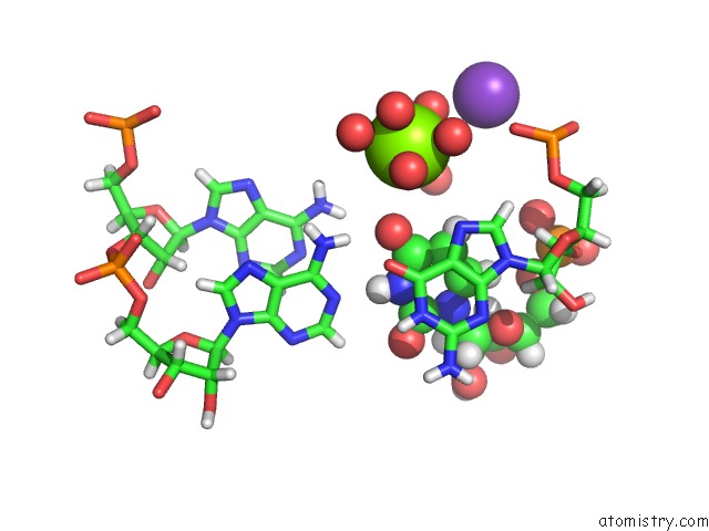
Magnesium Binding Sites:
The binding sites of Magnesium atom in the Crystal Structure of SPCAS9 Variant Vrer Bound to Sgrna and Tgcg Pam Target Dna
(pdb code 5fw3). This binding sites where shown within
5.0 Angstroms radius around Magnesium atom.
In total 2 binding sites of Magnesium where determined in the Crystal Structure of SPCAS9 Variant Vrer Bound to Sgrna and Tgcg Pam Target Dna, PDB code: 5fw3:
Jump to Magnesium binding site number: 1; 2;
In total 2 binding sites of Magnesium where determined in the Crystal Structure of SPCAS9 Variant Vrer Bound to Sgrna and Tgcg Pam Target Dna, PDB code: 5fw3:
Jump to Magnesium binding site number: 1; 2;
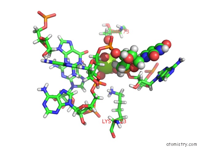
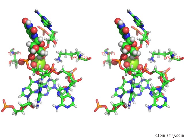
Magnesium binding site 1 out of 2 in 5fw3
Go back to
Magnesium binding site 1 out
of 2 in the Crystal Structure of SPCAS9 Variant Vrer Bound to Sgrna and Tgcg Pam Target Dna
Mono view
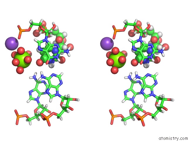
Stereo pair view
Mono view
Stereo pair view
A full contact list of Magnesium with other atoms in the Mg binding
site number 1 of Crystal Structure of SPCAS9 Variant Vrer Bound to Sgrna and Tgcg Pam Target Dna within 5.0Å range:
|
Magnesium binding site 2 out of 2 in 5fw3
Go back to
Magnesium binding site 2 out
of 2 in the Crystal Structure of SPCAS9 Variant Vrer Bound to Sgrna and Tgcg Pam Target Dna
Mono view
Stereo pair view
Mono view
Stereo pair view
A full contact list of Magnesium with other atoms in the Mg binding
site number 2 of Crystal Structure of SPCAS9 Variant Vrer Bound to Sgrna and Tgcg Pam Target Dna within 5.0Å range:
|
Reference:
C.Anders,
K.Bargsten,
M.Jinek.
Structural Plasticity of Pam Recognition By Engineered Variants of the Rna-Guided Endonuclease CAS9. Mol.Cell V. 61 895 2016.
ISSN: ISSN 1097-2765
PubMed: 26990992
DOI: 10.1016/J.MOLCEL.2016.02.020
Page generated: Sun Sep 29 04:32:32 2024
ISSN: ISSN 1097-2765
PubMed: 26990992
DOI: 10.1016/J.MOLCEL.2016.02.020
Last articles
Cl in 7U6HCl in 7U9A
Cl in 7U99
Cl in 7U56
Cl in 7U92
Cl in 7U68
Cl in 7U8E
Cl in 7U55
Cl in 7U5Z
Cl in 7U4K