Magnesium »
PDB 5grf-5h2f »
5gue »
Magnesium in PDB 5gue: Crystal Structure of COTB2 (Ggspp/MG2+-Bound Form) From Streptomyces Melanosporofaciens
Enzymatic activity of Crystal Structure of COTB2 (Ggspp/MG2+-Bound Form) From Streptomyces Melanosporofaciens
All present enzymatic activity of Crystal Structure of COTB2 (Ggspp/MG2+-Bound Form) From Streptomyces Melanosporofaciens:
4.2.3.146;
4.2.3.146;
Protein crystallography data
The structure of Crystal Structure of COTB2 (Ggspp/MG2+-Bound Form) From Streptomyces Melanosporofaciens, PDB code: 5gue
was solved by
T.Tomita,
S.-Y.Kim,
T.Ozaki,
A.Yoshida,
T.Kuzuyama,
M.Nishiyama,
with X-Ray Crystallography technique. A brief refinement statistics is given in the table below:
| Resolution Low / High (Å) | 34.15 / 1.80 |
| Space group | P 1 21 1 |
| Cell size a, b, c (Å), α, β, γ (°) | 58.698, 100.373, 108.937, 90.00, 90.00, 90.00 |
| R / Rfree (%) | 19.1 / 23.1 |
Magnesium Binding Sites:
The binding sites of Magnesium atom in the Crystal Structure of COTB2 (Ggspp/MG2+-Bound Form) From Streptomyces Melanosporofaciens
(pdb code 5gue). This binding sites where shown within
5.0 Angstroms radius around Magnesium atom.
In total 4 binding sites of Magnesium where determined in the Crystal Structure of COTB2 (Ggspp/MG2+-Bound Form) From Streptomyces Melanosporofaciens, PDB code: 5gue:
Jump to Magnesium binding site number: 1; 2; 3; 4;
In total 4 binding sites of Magnesium where determined in the Crystal Structure of COTB2 (Ggspp/MG2+-Bound Form) From Streptomyces Melanosporofaciens, PDB code: 5gue:
Jump to Magnesium binding site number: 1; 2; 3; 4;
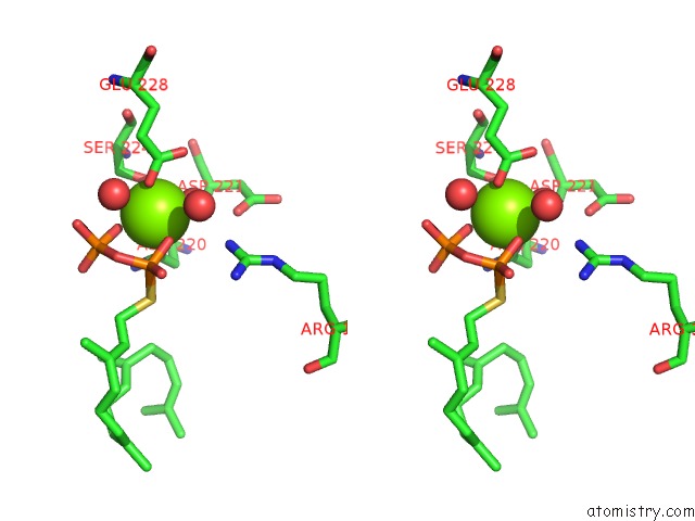
Magnesium binding site 1 out of 4 in 5gue
Go back to
Magnesium binding site 1 out
of 4 in the Crystal Structure of COTB2 (Ggspp/MG2+-Bound Form) From Streptomyces Melanosporofaciens

Mono view

Stereo pair view
Mono view
Stereo pair view
A full contact list of Magnesium with other atoms in the Mg binding
site number 1 of Crystal Structure of COTB2 (Ggspp/MG2+-Bound Form) From Streptomyces Melanosporofaciens within 5.0Å range:
|
Magnesium binding site 2 out of 4 in 5gue
Go back to
Magnesium binding site 2 out
of 4 in the Crystal Structure of COTB2 (Ggspp/MG2+-Bound Form) From Streptomyces Melanosporofaciens

Mono view

Stereo pair view
Mono view
Stereo pair view
A full contact list of Magnesium with other atoms in the Mg binding
site number 2 of Crystal Structure of COTB2 (Ggspp/MG2+-Bound Form) From Streptomyces Melanosporofaciens within 5.0Å range:
|
Magnesium binding site 3 out of 4 in 5gue
Go back to
Magnesium binding site 3 out
of 4 in the Crystal Structure of COTB2 (Ggspp/MG2+-Bound Form) From Streptomyces Melanosporofaciens

Mono view
Stereo pair view
Mono view
Stereo pair view
A full contact list of Magnesium with other atoms in the Mg binding
site number 3 of Crystal Structure of COTB2 (Ggspp/MG2+-Bound Form) From Streptomyces Melanosporofaciens within 5.0Å range:
|
Magnesium binding site 4 out of 4 in 5gue
Go back to
Magnesium binding site 4 out
of 4 in the Crystal Structure of COTB2 (Ggspp/MG2+-Bound Form) From Streptomyces Melanosporofaciens

Mono view

Stereo pair view
Mono view
Stereo pair view
A full contact list of Magnesium with other atoms in the Mg binding
site number 4 of Crystal Structure of COTB2 (Ggspp/MG2+-Bound Form) From Streptomyces Melanosporofaciens within 5.0Å range:
|
Reference:
T.Tomita,
S.-Y.Kim,
K.Teramoto,
A.Meguro,
T.Ozaki,
A.Yoshida,
Y.Motoyoshi,
N.Mori,
K.Ishigami,
H.Watanabe,
M.Nishiyama,
T.Kuzuyama.
Structural Insights Into the COTB2-Catalyzed Cyclization of Geranylgeranyl Diphosphate to the Diterpene Cyclooctat-9-En-7-Ol Acs Chem. Biol. V. 12 1621 2017.
ISSN: ESSN 1554-8937
PubMed: 28463490
DOI: 10.1021/ACSCHEMBIO.7B00154
Page generated: Sun Sep 29 15:26:06 2024
ISSN: ESSN 1554-8937
PubMed: 28463490
DOI: 10.1021/ACSCHEMBIO.7B00154
Last articles
Zn in 9MJ5Zn in 9HNW
Zn in 9G0L
Zn in 9FNE
Zn in 9DZN
Zn in 9E0I
Zn in 9D32
Zn in 9DAK
Zn in 8ZXC
Zn in 8ZUF