Magnesium »
PDB 5gre-5h1y »
5gx2 »
Magnesium in PDB 5gx2: Luciferin-Regenerating Enzyme Collected with Serial Synchrotron Rotational Crystallography with Accumulated Dose of 3.4 Mgy (3RD Measurement)
Protein crystallography data
The structure of Luciferin-Regenerating Enzyme Collected with Serial Synchrotron Rotational Crystallography with Accumulated Dose of 3.4 Mgy (3RD Measurement), PDB code: 5gx2
was solved by
K.Hasegawa,
K.Yamashita,
T.Murai,
N.Nuemket,
K.Hirata,
G.Ueno,
H.Ago,
T.Nakatsu,
T.Kumasaka,
M.Yamamoto,
with X-Ray Crystallography technique. A brief refinement statistics is given in the table below:
| Resolution Low / High (Å) | 42.13 / 1.60 |
| Space group | P 21 21 21 |
| Cell size a, b, c (Å), α, β, γ (°) | 47.441, 76.781, 84.263, 90.00, 90.00, 90.00 |
| R / Rfree (%) | 15.8 / 17.2 |
Other elements in 5gx2:
The structure of Luciferin-Regenerating Enzyme Collected with Serial Synchrotron Rotational Crystallography with Accumulated Dose of 3.4 Mgy (3RD Measurement) also contains other interesting chemical elements:
| Mercury | (Hg) | 2 atoms |
Magnesium Binding Sites:
The binding sites of Magnesium atom in the Luciferin-Regenerating Enzyme Collected with Serial Synchrotron Rotational Crystallography with Accumulated Dose of 3.4 Mgy (3RD Measurement)
(pdb code 5gx2). This binding sites where shown within
5.0 Angstroms radius around Magnesium atom.
In total only one binding site of Magnesium was determined in the Luciferin-Regenerating Enzyme Collected with Serial Synchrotron Rotational Crystallography with Accumulated Dose of 3.4 Mgy (3RD Measurement), PDB code: 5gx2:
In total only one binding site of Magnesium was determined in the Luciferin-Regenerating Enzyme Collected with Serial Synchrotron Rotational Crystallography with Accumulated Dose of 3.4 Mgy (3RD Measurement), PDB code: 5gx2:
Magnesium binding site 1 out of 1 in 5gx2
Go back to
Magnesium binding site 1 out
of 1 in the Luciferin-Regenerating Enzyme Collected with Serial Synchrotron Rotational Crystallography with Accumulated Dose of 3.4 Mgy (3RD Measurement)
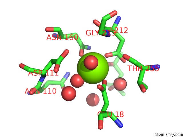
Mono view
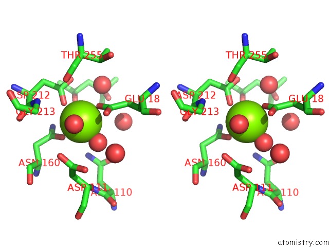
Stereo pair view
Mono view
Stereo pair view
A full contact list of Magnesium with other atoms in the Mg binding
site number 1 of Luciferin-Regenerating Enzyme Collected with Serial Synchrotron Rotational Crystallography with Accumulated Dose of 3.4 Mgy (3RD Measurement) within 5.0Å range:
|
Reference:
K.Hasegawa,
K.Yamashita,
T.Murai,
N.Nuemket,
K.Hirata,
G.Ueno,
H.Ago,
T.Nakatsu,
T.Kumasaka,
M.Yamamoto.
Development of A Dose-Limiting Data Collection Strategy For Serial Synchrotron Rotation Crystallography J Synchrotron Radiat V. 24 29 2017.
ISSN: ESSN 1600-5775
PubMed: 28009544
DOI: 10.1107/S1600577516016362
Page generated: Sun Sep 29 15:29:47 2024
ISSN: ESSN 1600-5775
PubMed: 28009544
DOI: 10.1107/S1600577516016362
Last articles
Zn in 9JYWZn in 9IR4
Zn in 9IR3
Zn in 9GMX
Zn in 9GMW
Zn in 9JEJ
Zn in 9ERF
Zn in 9ERE
Zn in 9EGV
Zn in 9EGW