Magnesium »
PDB 5h2q-5hia »
5h3f »
Magnesium in PDB 5h3f: Crystal Structure of Mouse Isocitrate Dehydrogenases 2 Complexed with Isocitrate
Enzymatic activity of Crystal Structure of Mouse Isocitrate Dehydrogenases 2 Complexed with Isocitrate
All present enzymatic activity of Crystal Structure of Mouse Isocitrate Dehydrogenases 2 Complexed with Isocitrate:
1.1.1.42;
1.1.1.42;
Protein crystallography data
The structure of Crystal Structure of Mouse Isocitrate Dehydrogenases 2 Complexed with Isocitrate, PDB code: 5h3f
was solved by
Y.Xu,
L.Liu,
T.Miyakawa,
A.Nakamura,
M.Tanokura,
with X-Ray Crystallography technique. A brief refinement statistics is given in the table below:
| Resolution Low / High (Å) | 19.89 / 3.29 |
| Space group | P 32 2 1 |
| Cell size a, b, c (Å), α, β, γ (°) | 135.853, 135.853, 220.162, 90.00, 90.00, 120.00 |
| R / Rfree (%) | 19.2 / 22.2 |
Magnesium Binding Sites:
The binding sites of Magnesium atom in the Crystal Structure of Mouse Isocitrate Dehydrogenases 2 Complexed with Isocitrate
(pdb code 5h3f). This binding sites where shown within
5.0 Angstroms radius around Magnesium atom.
In total 2 binding sites of Magnesium where determined in the Crystal Structure of Mouse Isocitrate Dehydrogenases 2 Complexed with Isocitrate, PDB code: 5h3f:
Jump to Magnesium binding site number: 1; 2;
In total 2 binding sites of Magnesium where determined in the Crystal Structure of Mouse Isocitrate Dehydrogenases 2 Complexed with Isocitrate, PDB code: 5h3f:
Jump to Magnesium binding site number: 1; 2;
Magnesium binding site 1 out of 2 in 5h3f
Go back to
Magnesium binding site 1 out
of 2 in the Crystal Structure of Mouse Isocitrate Dehydrogenases 2 Complexed with Isocitrate
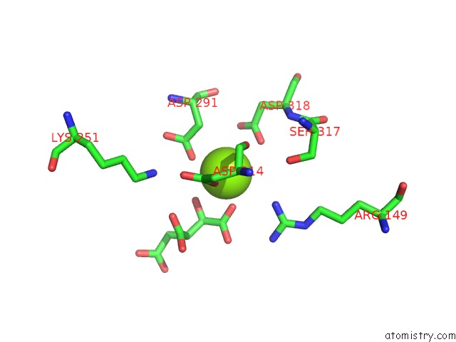
Mono view
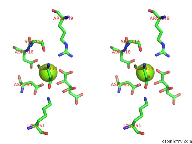
Stereo pair view
Mono view
Stereo pair view
A full contact list of Magnesium with other atoms in the Mg binding
site number 1 of Crystal Structure of Mouse Isocitrate Dehydrogenases 2 Complexed with Isocitrate within 5.0Å range:
|
Magnesium binding site 2 out of 2 in 5h3f
Go back to
Magnesium binding site 2 out
of 2 in the Crystal Structure of Mouse Isocitrate Dehydrogenases 2 Complexed with Isocitrate

Mono view
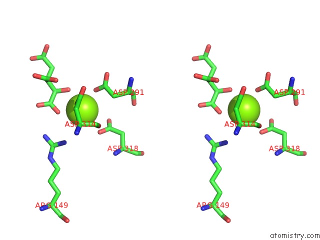
Stereo pair view
Mono view
Stereo pair view
A full contact list of Magnesium with other atoms in the Mg binding
site number 2 of Crystal Structure of Mouse Isocitrate Dehydrogenases 2 Complexed with Isocitrate within 5.0Å range:
|
Reference:
Y.Xu,
L.Liu,
A.Nakamura,
S.Someya,
T.Miyakawa,
M.Tanokura.
Studies on the Regulatory Mechanism of Isocitrate Dehydrogenase 2 Using Acetylation Mimics Sci Rep V. 7 9785 2017.
ISSN: ESSN 2045-2322
PubMed: 28852116
DOI: 10.1038/S41598-017-10337-7
Page generated: Sun Sep 29 15:40:54 2024
ISSN: ESSN 2045-2322
PubMed: 28852116
DOI: 10.1038/S41598-017-10337-7
Last articles
Fe in 2YXOFe in 2YRS
Fe in 2YXC
Fe in 2YNM
Fe in 2YVJ
Fe in 2YP1
Fe in 2YU2
Fe in 2YU1
Fe in 2YQB
Fe in 2YOO