Magnesium »
PDB 5h2q-5hia »
5h8u »
Magnesium in PDB 5h8u: Crystal Structure of Mycobacterium Tuberculosis Wild-Type Malate Synthase in Complex with Product Malate
Enzymatic activity of Crystal Structure of Mycobacterium Tuberculosis Wild-Type Malate Synthase in Complex with Product Malate
All present enzymatic activity of Crystal Structure of Mycobacterium Tuberculosis Wild-Type Malate Synthase in Complex with Product Malate:
2.3.3.9;
2.3.3.9;
Protein crystallography data
The structure of Crystal Structure of Mycobacterium Tuberculosis Wild-Type Malate Synthase in Complex with Product Malate, PDB code: 5h8u
was solved by
I.V.Krieger,
H.-L.Huang,
J.C.Sacchettini,
with X-Ray Crystallography technique. A brief refinement statistics is given in the table below:
| Resolution Low / High (Å) | 37.36 / 2.85 |
| Space group | P 41 21 2 |
| Cell size a, b, c (Å), α, β, γ (°) | 120.659, 120.659, 232.224, 90.00, 90.00, 90.00 |
| R / Rfree (%) | 22.6 / 27.6 |
Magnesium Binding Sites:
The binding sites of Magnesium atom in the Crystal Structure of Mycobacterium Tuberculosis Wild-Type Malate Synthase in Complex with Product Malate
(pdb code 5h8u). This binding sites where shown within
5.0 Angstroms radius around Magnesium atom.
In total 3 binding sites of Magnesium where determined in the Crystal Structure of Mycobacterium Tuberculosis Wild-Type Malate Synthase in Complex with Product Malate, PDB code: 5h8u:
Jump to Magnesium binding site number: 1; 2; 3;
In total 3 binding sites of Magnesium where determined in the Crystal Structure of Mycobacterium Tuberculosis Wild-Type Malate Synthase in Complex with Product Malate, PDB code: 5h8u:
Jump to Magnesium binding site number: 1; 2; 3;
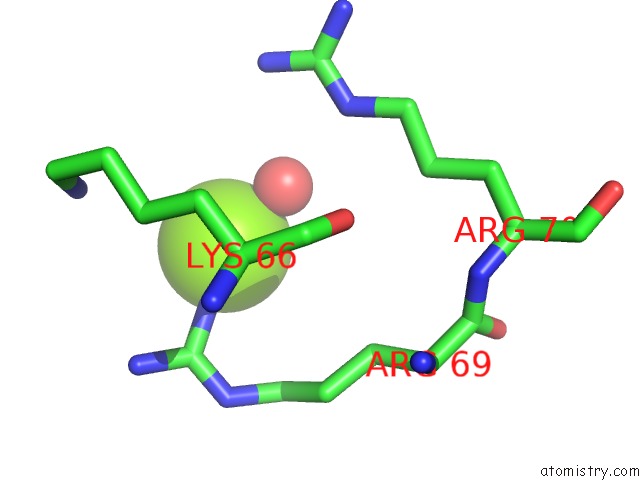
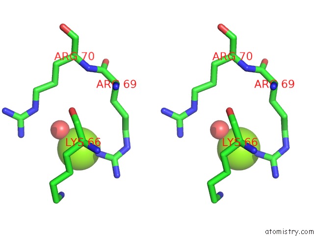
Magnesium binding site 1 out of 3 in 5h8u
Go back to
Magnesium binding site 1 out
of 3 in the Crystal Structure of Mycobacterium Tuberculosis Wild-Type Malate Synthase in Complex with Product Malate
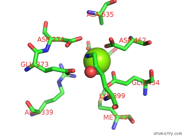
Mono view
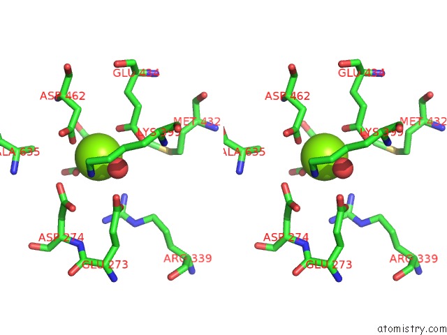
Stereo pair view
Mono view
Stereo pair view
A full contact list of Magnesium with other atoms in the Mg binding
site number 1 of Crystal Structure of Mycobacterium Tuberculosis Wild-Type Malate Synthase in Complex with Product Malate within 5.0Å range:
|
Magnesium binding site 2 out of 3 in 5h8u
Go back to
Magnesium binding site 2 out
of 3 in the Crystal Structure of Mycobacterium Tuberculosis Wild-Type Malate Synthase in Complex with Product Malate
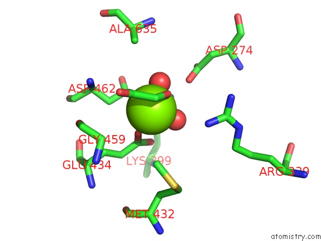
Mono view
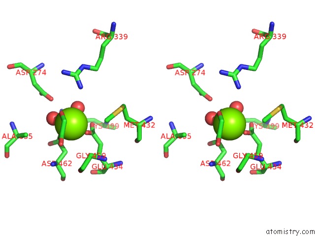
Stereo pair view
Mono view
Stereo pair view
A full contact list of Magnesium with other atoms in the Mg binding
site number 2 of Crystal Structure of Mycobacterium Tuberculosis Wild-Type Malate Synthase in Complex with Product Malate within 5.0Å range:
|
Magnesium binding site 3 out of 3 in 5h8u
Go back to
Magnesium binding site 3 out
of 3 in the Crystal Structure of Mycobacterium Tuberculosis Wild-Type Malate Synthase in Complex with Product Malate
Mono view
Stereo pair view
Mono view
Stereo pair view
A full contact list of Magnesium with other atoms in the Mg binding
site number 3 of Crystal Structure of Mycobacterium Tuberculosis Wild-Type Malate Synthase in Complex with Product Malate within 5.0Å range:
|
Reference:
H.L.Huang,
I.V.Krieger,
M.K.Parai,
V.B.Gawandi,
J.C.Sacchettini.
Mycobacterium Tuberculosis Malate Synthase Structures with Fragments Reveal A Portal For Substrate/Product Exchange. J. Biol. Chem. V. 291 27421 2016.
ISSN: ESSN 1083-351X
PubMed: 27738104
DOI: 10.1074/JBC.M116.750877
Page generated: Tue Aug 12 10:43:50 2025
ISSN: ESSN 1083-351X
PubMed: 27738104
DOI: 10.1074/JBC.M116.750877
Last articles
Mg in 6IAHMg in 6IB7
Mg in 6IAN
Mg in 6I9P
Mg in 6I9T
Mg in 6IAF
Mg in 6IAE
Mg in 6IA7
Mg in 6I8F
Mg in 6I5X