Magnesium »
PDB 5h2f-5hi8 »
5hi8 »
Magnesium in PDB 5hi8: Structure of T-Type Phycobiliprotein Lyase Cpet From Prochlorococcus Phage P-HM1
Protein crystallography data
The structure of Structure of T-Type Phycobiliprotein Lyase Cpet From Prochlorococcus Phage P-HM1, PDB code: 5hi8
was solved by
R.Gasper,
J.Schwach,
N.Frankenberg-Dinkel,
E.Hofmann,
with X-Ray Crystallography technique. A brief refinement statistics is given in the table below:
| Resolution Low / High (Å) | 43.92 / 1.80 |
| Space group | C 1 2 1 |
| Cell size a, b, c (Å), α, β, γ (°) | 63.290, 61.680, 93.340, 90.00, 109.77, 90.00 |
| R / Rfree (%) | 20.7 / 22.8 |
Magnesium Binding Sites:
The binding sites of Magnesium atom in the Structure of T-Type Phycobiliprotein Lyase Cpet From Prochlorococcus Phage P-HM1
(pdb code 5hi8). This binding sites where shown within
5.0 Angstroms radius around Magnesium atom.
In total 2 binding sites of Magnesium where determined in the Structure of T-Type Phycobiliprotein Lyase Cpet From Prochlorococcus Phage P-HM1, PDB code: 5hi8:
Jump to Magnesium binding site number: 1; 2;
In total 2 binding sites of Magnesium where determined in the Structure of T-Type Phycobiliprotein Lyase Cpet From Prochlorococcus Phage P-HM1, PDB code: 5hi8:
Jump to Magnesium binding site number: 1; 2;
Magnesium binding site 1 out of 2 in 5hi8
Go back to
Magnesium binding site 1 out
of 2 in the Structure of T-Type Phycobiliprotein Lyase Cpet From Prochlorococcus Phage P-HM1
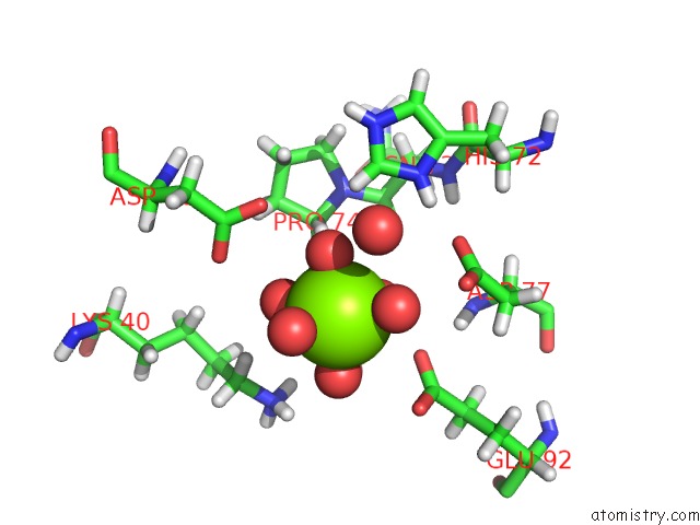
Mono view
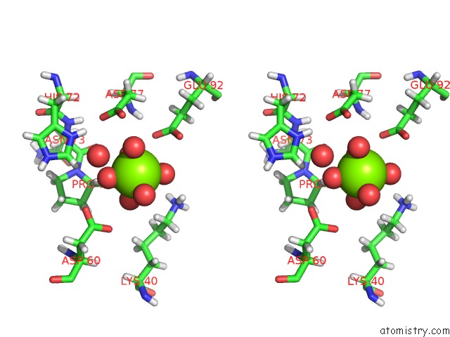
Stereo pair view
Mono view
Stereo pair view
A full contact list of Magnesium with other atoms in the Mg binding
site number 1 of Structure of T-Type Phycobiliprotein Lyase Cpet From Prochlorococcus Phage P-HM1 within 5.0Å range:
|
Magnesium binding site 2 out of 2 in 5hi8
Go back to
Magnesium binding site 2 out
of 2 in the Structure of T-Type Phycobiliprotein Lyase Cpet From Prochlorococcus Phage P-HM1
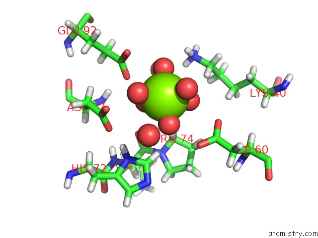
Mono view
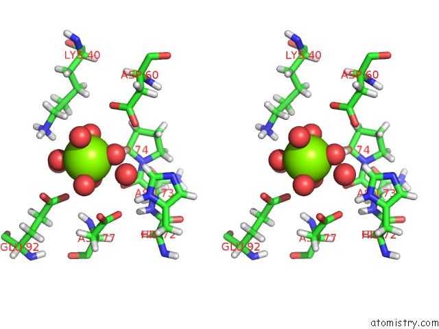
Stereo pair view
Mono view
Stereo pair view
A full contact list of Magnesium with other atoms in the Mg binding
site number 2 of Structure of T-Type Phycobiliprotein Lyase Cpet From Prochlorococcus Phage P-HM1 within 5.0Å range:
|
Reference:
R.Gasper,
J.Schwach,
J.Hartmann,
A.Holtkamp,
J.Wiethaus,
N.Riedel,
E.Hofmann,
N.Frankenberg-Dinkel.
Distinct Features of Cyanophage-Encoded T-Type Phycobiliprotein Lyase Phi Cpet: the Role of Auxiliary Metabolic Genes. J. Biol. Chem. V. 292 3089 2017.
ISSN: ESSN 1083-351X
PubMed: 28073912
DOI: 10.1074/JBC.M116.769703
Page generated: Sun Sep 29 15:49:38 2024
ISSN: ESSN 1083-351X
PubMed: 28073912
DOI: 10.1074/JBC.M116.769703
Last articles
Zn in 9J0NZn in 9J0O
Zn in 9J0P
Zn in 9FJX
Zn in 9EKB
Zn in 9C0F
Zn in 9CAH
Zn in 9CH0
Zn in 9CH3
Zn in 9CH1