Magnesium »
PDB 5hia-5hqw »
5hml »
Magnesium in PDB 5hml: Crystal Structure of T5 D15 Protein Co-Crystallized with Metal Ions
Enzymatic activity of Crystal Structure of T5 D15 Protein Co-Crystallized with Metal Ions
All present enzymatic activity of Crystal Structure of T5 D15 Protein Co-Crystallized with Metal Ions:
3.1.11.3;
3.1.11.3;
Protein crystallography data
The structure of Crystal Structure of T5 D15 Protein Co-Crystallized with Metal Ions, PDB code: 5hml
was solved by
C.S.Flemming,
M.Feng,
S.E.Sedelnikova,
J.Zhang,
J.B.Rafferty,
J.R.Sayers,
P.J.Artymiuk,
with X-Ray Crystallography technique. A brief refinement statistics is given in the table below:
| Resolution Low / High (Å) | 54.60 / 1.48 |
| Space group | P 1 |
| Cell size a, b, c (Å), α, β, γ (°) | 47.380, 58.380, 59.720, 66.53, 79.48, 73.71 |
| R / Rfree (%) | 14.1 / 19.6 |
Other elements in 5hml:
The structure of Crystal Structure of T5 D15 Protein Co-Crystallized with Metal Ions also contains other interesting chemical elements:
| Iodine | (I) | 3 atoms |
| Chlorine | (Cl) | 13 atoms |
Magnesium Binding Sites:
The binding sites of Magnesium atom in the Crystal Structure of T5 D15 Protein Co-Crystallized with Metal Ions
(pdb code 5hml). This binding sites where shown within
5.0 Angstroms radius around Magnesium atom.
In total 3 binding sites of Magnesium where determined in the Crystal Structure of T5 D15 Protein Co-Crystallized with Metal Ions, PDB code: 5hml:
Jump to Magnesium binding site number: 1; 2; 3;
In total 3 binding sites of Magnesium where determined in the Crystal Structure of T5 D15 Protein Co-Crystallized with Metal Ions, PDB code: 5hml:
Jump to Magnesium binding site number: 1; 2; 3;
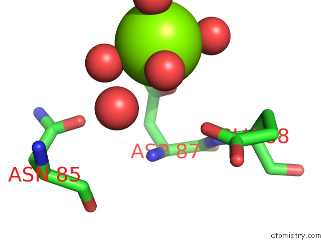
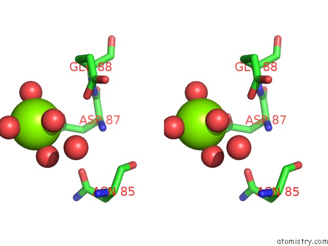
Magnesium binding site 1 out of 3 in 5hml
Go back to
Magnesium binding site 1 out
of 3 in the Crystal Structure of T5 D15 Protein Co-Crystallized with Metal Ions
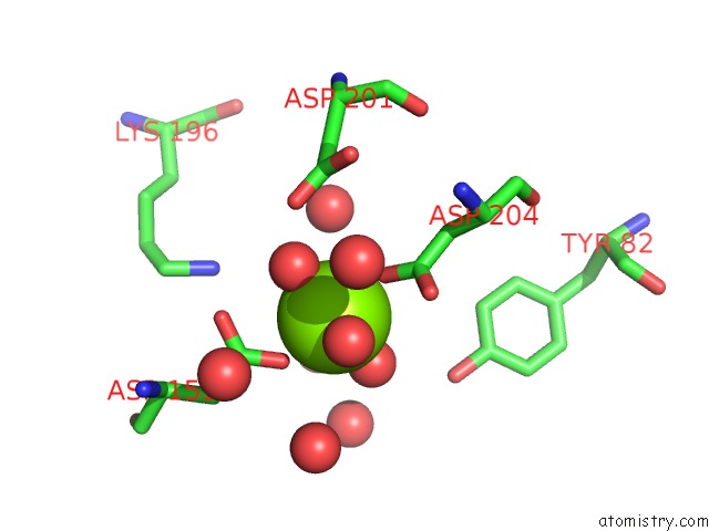
Mono view
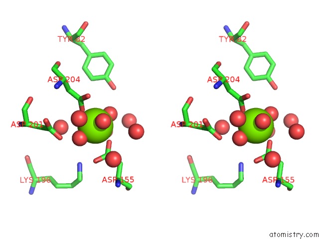
Stereo pair view
Mono view
Stereo pair view
A full contact list of Magnesium with other atoms in the Mg binding
site number 1 of Crystal Structure of T5 D15 Protein Co-Crystallized with Metal Ions within 5.0Å range:
|
Magnesium binding site 2 out of 3 in 5hml
Go back to
Magnesium binding site 2 out
of 3 in the Crystal Structure of T5 D15 Protein Co-Crystallized with Metal Ions
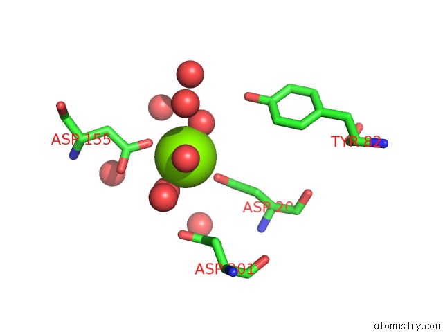
Mono view
Stereo pair view
Mono view
Stereo pair view
A full contact list of Magnesium with other atoms in the Mg binding
site number 2 of Crystal Structure of T5 D15 Protein Co-Crystallized with Metal Ions within 5.0Å range:
|
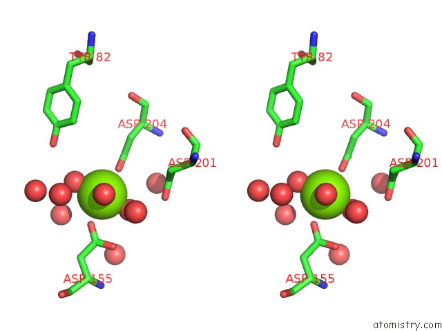
Magnesium binding site 3 out of 3 in 5hml
Go back to
Magnesium binding site 3 out
of 3 in the Crystal Structure of T5 D15 Protein Co-Crystallized with Metal Ions
Mono view
Stereo pair view
Mono view
Stereo pair view
A full contact list of Magnesium with other atoms in the Mg binding
site number 3 of Crystal Structure of T5 D15 Protein Co-Crystallized with Metal Ions within 5.0Å range:
|
Reference:
F.A.Almalki,
C.S.Flemming,
J.Zhang,
M.Feng,
S.E.Sedelnikova,
T.Ceska,
J.B.Rafferty,
J.R.Sayers,
P.J.Artymiuk.
Direct Observation of Dna Threading in Flap Endonuclease Complexes. Nat.Struct.Mol.Biol. V. 23 640 2016.
ISSN: ESSN 1545-9985
PubMed: 27273516
DOI: 10.1038/NSMB.3241
Page generated: Sun Sep 29 16:01:02 2024
ISSN: ESSN 1545-9985
PubMed: 27273516
DOI: 10.1038/NSMB.3241
Last articles
Zn in 9J0NZn in 9J0O
Zn in 9J0P
Zn in 9FJX
Zn in 9EKB
Zn in 9C0F
Zn in 9CAH
Zn in 9CH0
Zn in 9CH3
Zn in 9CH1