Magnesium »
PDB 5hia-5hqw »
5hqb »
Magnesium in PDB 5hqb: A Glycoside Hydrolase Family 97 Enzyme (E480Q) in Complex with Panose From Pseudoalteromonas Sp. Strain K8
Protein crystallography data
The structure of A Glycoside Hydrolase Family 97 Enzyme (E480Q) in Complex with Panose From Pseudoalteromonas Sp. Strain K8, PDB code: 5hqb
was solved by
J.Li,
C.He,
Y.Xiao,
with X-Ray Crystallography technique. A brief refinement statistics is given in the table below:
| Resolution Low / High (Å) | 39.29 / 1.80 |
| Space group | P 32 2 1 |
| Cell size a, b, c (Å), α, β, γ (°) | 119.167, 119.167, 181.832, 90.00, 90.00, 120.00 |
| R / Rfree (%) | 13.7 / 15.5 |
Other elements in 5hqb:
The structure of A Glycoside Hydrolase Family 97 Enzyme (E480Q) in Complex with Panose From Pseudoalteromonas Sp. Strain K8 also contains other interesting chemical elements:
| Calcium | (Ca) | 1 atom |
| Chlorine | (Cl) | 2 atoms |
Magnesium Binding Sites:
The binding sites of Magnesium atom in the A Glycoside Hydrolase Family 97 Enzyme (E480Q) in Complex with Panose From Pseudoalteromonas Sp. Strain K8
(pdb code 5hqb). This binding sites where shown within
5.0 Angstroms radius around Magnesium atom.
In total only one binding site of Magnesium was determined in the A Glycoside Hydrolase Family 97 Enzyme (E480Q) in Complex with Panose From Pseudoalteromonas Sp. Strain K8, PDB code: 5hqb:
In total only one binding site of Magnesium was determined in the A Glycoside Hydrolase Family 97 Enzyme (E480Q) in Complex with Panose From Pseudoalteromonas Sp. Strain K8, PDB code: 5hqb:
Magnesium binding site 1 out of 1 in 5hqb
Go back to
Magnesium binding site 1 out
of 1 in the A Glycoside Hydrolase Family 97 Enzyme (E480Q) in Complex with Panose From Pseudoalteromonas Sp. Strain K8
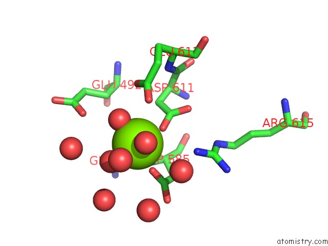
Mono view
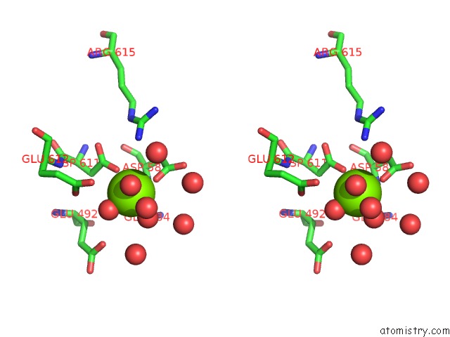
Stereo pair view
Mono view
Stereo pair view
A full contact list of Magnesium with other atoms in the Mg binding
site number 1 of A Glycoside Hydrolase Family 97 Enzyme (E480Q) in Complex with Panose From Pseudoalteromonas Sp. Strain K8 within 5.0Å range:
|
Reference:
C.He,
J.Li,
W.Li,
Y.Xue,
Z.Fang,
W.Fang,
X.Zhang,
X.Wang,
Y.Xiao.
Structures of PSPAG97A Alpha-Glucoside Hydrolase Reveal A Novel Mechanism For Chloride Induced Activation. J. Struct. Biol. V. 196 426 2016.
ISSN: ESSN 1095-8657
PubMed: 27645700
DOI: 10.1016/J.JSB.2016.09.009
Page generated: Sun Sep 29 16:06:15 2024
ISSN: ESSN 1095-8657
PubMed: 27645700
DOI: 10.1016/J.JSB.2016.09.009
Last articles
Zn in 9J0NZn in 9J0O
Zn in 9J0P
Zn in 9FJX
Zn in 9EKB
Zn in 9C0F
Zn in 9CAH
Zn in 9CH0
Zn in 9CH3
Zn in 9CH1