Magnesium »
PDB 5hr6-5i8q »
5hvk »
Magnesium in PDB 5hvk: Crystal Structure of LIMK1 Mutant D460N in Complex with Full-Length Cofilin-1
Enzymatic activity of Crystal Structure of LIMK1 Mutant D460N in Complex with Full-Length Cofilin-1
All present enzymatic activity of Crystal Structure of LIMK1 Mutant D460N in Complex with Full-Length Cofilin-1:
2.7.11.1;
2.7.11.1;
Protein crystallography data
The structure of Crystal Structure of LIMK1 Mutant D460N in Complex with Full-Length Cofilin-1, PDB code: 5hvk
was solved by
S.Hamill,
T.J.Boggon,
with X-Ray Crystallography technique. A brief refinement statistics is given in the table below:
| Resolution Low / High (Å) | 48.11 / 3.50 |
| Space group | P 21 21 21 |
| Cell size a, b, c (Å), α, β, γ (°) | 81.116, 102.283, 141.900, 90.00, 90.00, 90.00 |
| R / Rfree (%) | 27.3 / 31 |
Magnesium Binding Sites:
The binding sites of Magnesium atom in the Crystal Structure of LIMK1 Mutant D460N in Complex with Full-Length Cofilin-1
(pdb code 5hvk). This binding sites where shown within
5.0 Angstroms radius around Magnesium atom.
In total 2 binding sites of Magnesium where determined in the Crystal Structure of LIMK1 Mutant D460N in Complex with Full-Length Cofilin-1, PDB code: 5hvk:
Jump to Magnesium binding site number: 1; 2;
In total 2 binding sites of Magnesium where determined in the Crystal Structure of LIMK1 Mutant D460N in Complex with Full-Length Cofilin-1, PDB code: 5hvk:
Jump to Magnesium binding site number: 1; 2;
Magnesium binding site 1 out of 2 in 5hvk
Go back to
Magnesium binding site 1 out
of 2 in the Crystal Structure of LIMK1 Mutant D460N in Complex with Full-Length Cofilin-1
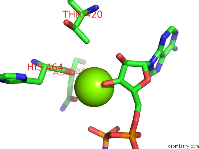
Mono view
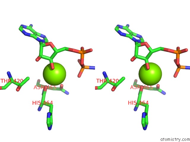
Stereo pair view
Mono view
Stereo pair view
A full contact list of Magnesium with other atoms in the Mg binding
site number 1 of Crystal Structure of LIMK1 Mutant D460N in Complex with Full-Length Cofilin-1 within 5.0Å range:
|
Magnesium binding site 2 out of 2 in 5hvk
Go back to
Magnesium binding site 2 out
of 2 in the Crystal Structure of LIMK1 Mutant D460N in Complex with Full-Length Cofilin-1
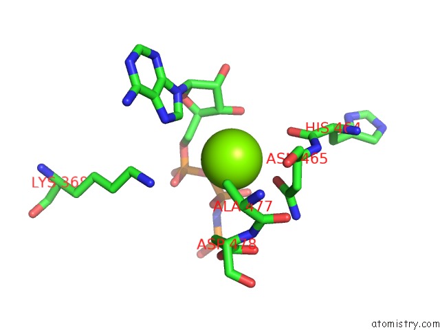
Mono view
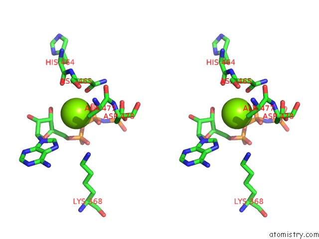
Stereo pair view
Mono view
Stereo pair view
A full contact list of Magnesium with other atoms in the Mg binding
site number 2 of Crystal Structure of LIMK1 Mutant D460N in Complex with Full-Length Cofilin-1 within 5.0Å range:
|
Reference:
S.Hamill,
H.J.Lou,
B.E.Turk,
T.J.Boggon.
Structural Basis For Noncanonical Substrate Recognition of Cofilin/Adf Proteins By Lim Kinases. Mol.Cell V. 62 397 2016.
ISSN: ISSN 1097-2765
PubMed: 27153537
DOI: 10.1016/J.MOLCEL.2016.04.001
Page generated: Sun Sep 29 16:35:50 2024
ISSN: ISSN 1097-2765
PubMed: 27153537
DOI: 10.1016/J.MOLCEL.2016.04.001
Last articles
Zn in 9J0NZn in 9J0O
Zn in 9J0P
Zn in 9FJX
Zn in 9EKB
Zn in 9C0F
Zn in 9CAH
Zn in 9CH0
Zn in 9CH3
Zn in 9CH1