Magnesium »
PDB 5ims-5iwa »
5it9 »
Magnesium in PDB 5it9: Structure of the Yeast Kluyveromyces Lactis Small Ribosomal Subunit in Complex with the Cricket Paralysis Virus Ires.
Other elements in 5it9:
The structure of Structure of the Yeast Kluyveromyces Lactis Small Ribosomal Subunit in Complex with the Cricket Paralysis Virus Ires. also contains other interesting chemical elements:
| Zinc | (Zn) | 3 atoms |
Magnesium Binding Sites:
Pages:
>>> Page 1 <<< Page 2, Binding sites: 11 - 20; Page 3, Binding sites: 21 - 30; Page 4, Binding sites: 31 - 40; Page 5, Binding sites: 41 - 50; Page 6, Binding sites: 51 - 60; Page 7, Binding sites: 61 - 70; Page 8, Binding sites: 71 - 80;Binding sites:
The binding sites of Magnesium atom in the Structure of the Yeast Kluyveromyces Lactis Small Ribosomal Subunit in Complex with the Cricket Paralysis Virus Ires. (pdb code 5it9). This binding sites where shown within 5.0 Angstroms radius around Magnesium atom.In total 80 binding sites of Magnesium where determined in the Structure of the Yeast Kluyveromyces Lactis Small Ribosomal Subunit in Complex with the Cricket Paralysis Virus Ires., PDB code: 5it9:
Jump to Magnesium binding site number: 1; 2; 3; 4; 5; 6; 7; 8; 9; 10;
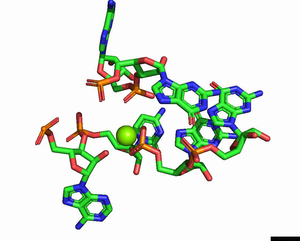
Magnesium binding site 1 out of 80 in 5it9
Go back to
Magnesium binding site 1 out
of 80 in the Structure of the Yeast Kluyveromyces Lactis Small Ribosomal Subunit in Complex with the Cricket Paralysis Virus Ires.

Mono view

Stereo pair view
Mono view
Stereo pair view
A full contact list of Magnesium with other atoms in the Mg binding
site number 1 of Structure of the Yeast Kluyveromyces Lactis Small Ribosomal Subunit in Complex with the Cricket Paralysis Virus Ires. within 5.0Å range:
|
Magnesium binding site 2 out of 80 in 5it9
Go back to
Magnesium binding site 2 out
of 80 in the Structure of the Yeast Kluyveromyces Lactis Small Ribosomal Subunit in Complex with the Cricket Paralysis Virus Ires.

Mono view

Stereo pair view
Mono view
Stereo pair view
A full contact list of Magnesium with other atoms in the Mg binding
site number 2 of Structure of the Yeast Kluyveromyces Lactis Small Ribosomal Subunit in Complex with the Cricket Paralysis Virus Ires. within 5.0Å range:
|
Magnesium binding site 3 out of 80 in 5it9
Go back to
Magnesium binding site 3 out
of 80 in the Structure of the Yeast Kluyveromyces Lactis Small Ribosomal Subunit in Complex with the Cricket Paralysis Virus Ires.

Mono view

Stereo pair view
Mono view
Stereo pair view
A full contact list of Magnesium with other atoms in the Mg binding
site number 3 of Structure of the Yeast Kluyveromyces Lactis Small Ribosomal Subunit in Complex with the Cricket Paralysis Virus Ires. within 5.0Å range:
|
Magnesium binding site 4 out of 80 in 5it9
Go back to
Magnesium binding site 4 out
of 80 in the Structure of the Yeast Kluyveromyces Lactis Small Ribosomal Subunit in Complex with the Cricket Paralysis Virus Ires.

Mono view

Stereo pair view
Mono view
Stereo pair view
A full contact list of Magnesium with other atoms in the Mg binding
site number 4 of Structure of the Yeast Kluyveromyces Lactis Small Ribosomal Subunit in Complex with the Cricket Paralysis Virus Ires. within 5.0Å range:
|
Magnesium binding site 5 out of 80 in 5it9
Go back to
Magnesium binding site 5 out
of 80 in the Structure of the Yeast Kluyveromyces Lactis Small Ribosomal Subunit in Complex with the Cricket Paralysis Virus Ires.

Mono view

Stereo pair view
Mono view
Stereo pair view
A full contact list of Magnesium with other atoms in the Mg binding
site number 5 of Structure of the Yeast Kluyveromyces Lactis Small Ribosomal Subunit in Complex with the Cricket Paralysis Virus Ires. within 5.0Å range:
|
Magnesium binding site 6 out of 80 in 5it9
Go back to
Magnesium binding site 6 out
of 80 in the Structure of the Yeast Kluyveromyces Lactis Small Ribosomal Subunit in Complex with the Cricket Paralysis Virus Ires.
Mono view

Stereo pair view
Mono view
Stereo pair view
A full contact list of Magnesium with other atoms in the Mg binding
site number 6 of Structure of the Yeast Kluyveromyces Lactis Small Ribosomal Subunit in Complex with the Cricket Paralysis Virus Ires. within 5.0Å range:
|
Magnesium binding site 7 out of 80 in 5it9
Go back to
Magnesium binding site 7 out
of 80 in the Structure of the Yeast Kluyveromyces Lactis Small Ribosomal Subunit in Complex with the Cricket Paralysis Virus Ires.

Mono view

Stereo pair view
Mono view
Stereo pair view
A full contact list of Magnesium with other atoms in the Mg binding
site number 7 of Structure of the Yeast Kluyveromyces Lactis Small Ribosomal Subunit in Complex with the Cricket Paralysis Virus Ires. within 5.0Å range:
|
Magnesium binding site 8 out of 80 in 5it9
Go back to
Magnesium binding site 8 out
of 80 in the Structure of the Yeast Kluyveromyces Lactis Small Ribosomal Subunit in Complex with the Cricket Paralysis Virus Ires.

Mono view

Stereo pair view
Mono view
Stereo pair view
A full contact list of Magnesium with other atoms in the Mg binding
site number 8 of Structure of the Yeast Kluyveromyces Lactis Small Ribosomal Subunit in Complex with the Cricket Paralysis Virus Ires. within 5.0Å range:
|
Magnesium binding site 9 out of 80 in 5it9
Go back to
Magnesium binding site 9 out
of 80 in the Structure of the Yeast Kluyveromyces Lactis Small Ribosomal Subunit in Complex with the Cricket Paralysis Virus Ires.

Mono view

Stereo pair view
Mono view
Stereo pair view
A full contact list of Magnesium with other atoms in the Mg binding
site number 9 of Structure of the Yeast Kluyveromyces Lactis Small Ribosomal Subunit in Complex with the Cricket Paralysis Virus Ires. within 5.0Å range:
|
Magnesium binding site 10 out of 80 in 5it9
Go back to
Magnesium binding site 10 out
of 80 in the Structure of the Yeast Kluyveromyces Lactis Small Ribosomal Subunit in Complex with the Cricket Paralysis Virus Ires.

Mono view

Stereo pair view
Mono view
Stereo pair view
A full contact list of Magnesium with other atoms in the Mg binding
site number 10 of Structure of the Yeast Kluyveromyces Lactis Small Ribosomal Subunit in Complex with the Cricket Paralysis Virus Ires. within 5.0Å range:
|
Reference:
J.Murray,
C.G.Savva,
B.S.Shin,
T.E.Dever,
V.Ramakrishnan,
I.S.Fernandez.
Structural Characterization of Ribosome Recruitment and Translocation By Type IV Ires. Elife V. 5 2016.
ISSN: ESSN 2050-084X
PubMed: 27159451
DOI: 10.7554/ELIFE.13567
Page generated: Sun Sep 29 16:56:14 2024
ISSN: ESSN 2050-084X
PubMed: 27159451
DOI: 10.7554/ELIFE.13567
Last articles
Zn in 9MJ5Zn in 9HNW
Zn in 9G0L
Zn in 9FNE
Zn in 9DZN
Zn in 9E0I
Zn in 9D32
Zn in 9DAK
Zn in 8ZXC
Zn in 8ZUF