Magnesium »
PDB 5ims-5iwa »
5iuj »
Magnesium in PDB 5iuj: Crystal Structure of the Desk-Desr Complex in the Phosphotransfer State with Low MG2+ (20 Mm)
Enzymatic activity of Crystal Structure of the Desk-Desr Complex in the Phosphotransfer State with Low MG2+ (20 Mm)
All present enzymatic activity of Crystal Structure of the Desk-Desr Complex in the Phosphotransfer State with Low MG2+ (20 Mm):
2.7.13.3;
2.7.13.3;
Protein crystallography data
The structure of Crystal Structure of the Desk-Desr Complex in the Phosphotransfer State with Low MG2+ (20 Mm), PDB code: 5iuj
was solved by
F.Trajtenberg,
J.A.Imelio,
N.Larrieux,
A.Buschiazzo,
with X-Ray Crystallography technique. A brief refinement statistics is given in the table below:
| Resolution Low / High (Å) | 66.70 / 3.20 |
| Space group | P 1 21 1 |
| Cell size a, b, c (Å), α, β, γ (°) | 87.820, 114.619, 91.597, 90.00, 116.44, 90.00 |
| R / Rfree (%) | 18.7 / 24 |
Other elements in 5iuj:
The structure of Crystal Structure of the Desk-Desr Complex in the Phosphotransfer State with Low MG2+ (20 Mm) also contains other interesting chemical elements:
| Potassium | (K) | 2 atoms |
Magnesium Binding Sites:
The binding sites of Magnesium atom in the Crystal Structure of the Desk-Desr Complex in the Phosphotransfer State with Low MG2+ (20 Mm)
(pdb code 5iuj). This binding sites where shown within
5.0 Angstroms radius around Magnesium atom.
In total 4 binding sites of Magnesium where determined in the Crystal Structure of the Desk-Desr Complex in the Phosphotransfer State with Low MG2+ (20 Mm), PDB code: 5iuj:
Jump to Magnesium binding site number: 1; 2; 3; 4;
In total 4 binding sites of Magnesium where determined in the Crystal Structure of the Desk-Desr Complex in the Phosphotransfer State with Low MG2+ (20 Mm), PDB code: 5iuj:
Jump to Magnesium binding site number: 1; 2; 3; 4;
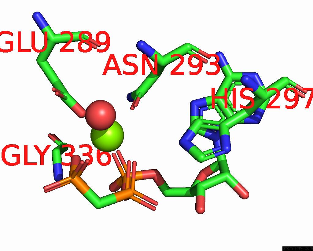
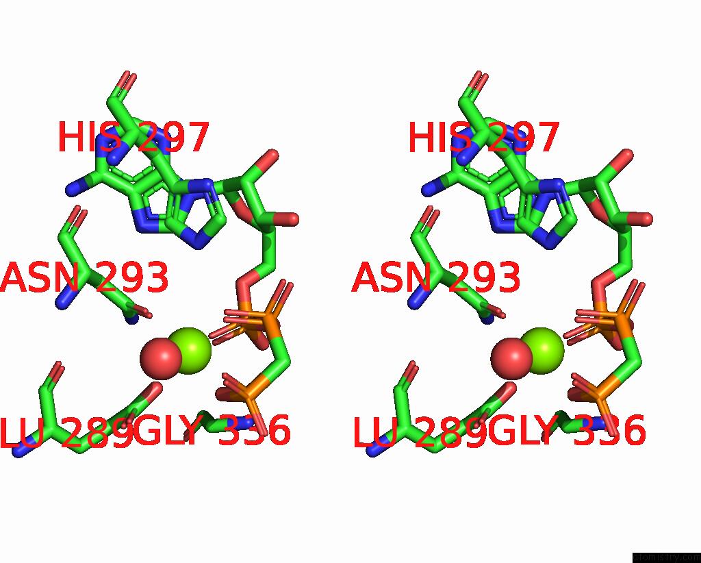
Magnesium binding site 1 out of 4 in 5iuj
Go back to
Magnesium binding site 1 out
of 4 in the Crystal Structure of the Desk-Desr Complex in the Phosphotransfer State with Low MG2+ (20 Mm)
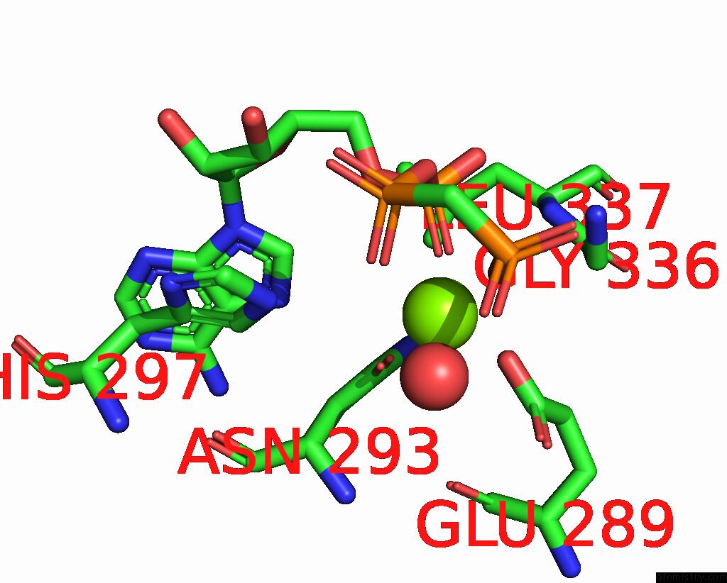
Mono view
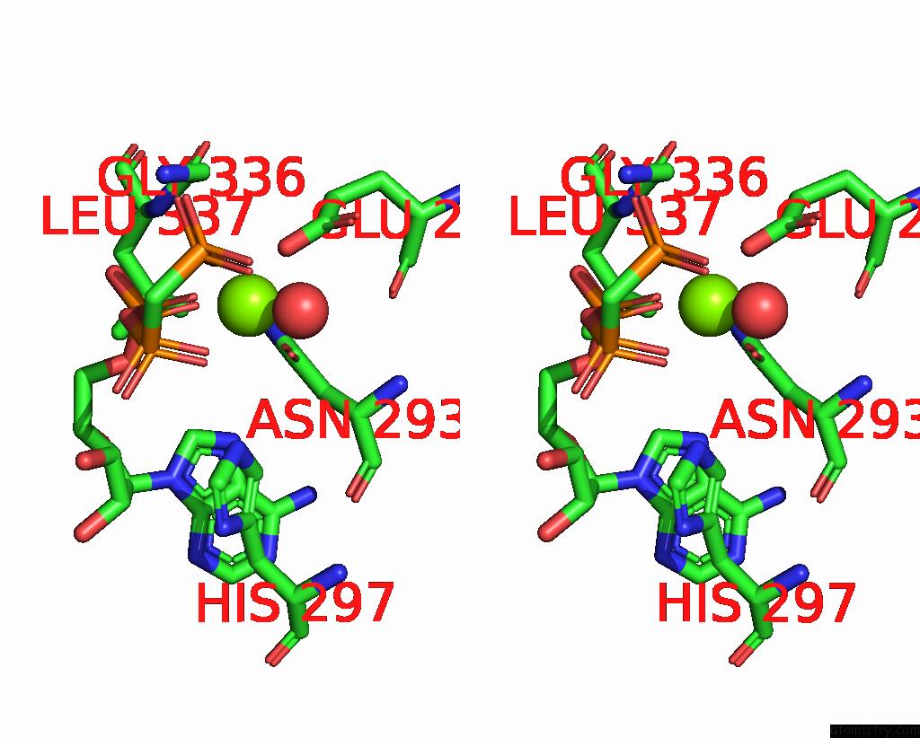
Stereo pair view
Mono view
Stereo pair view
A full contact list of Magnesium with other atoms in the Mg binding
site number 1 of Crystal Structure of the Desk-Desr Complex in the Phosphotransfer State with Low MG2+ (20 Mm) within 5.0Å range:
|
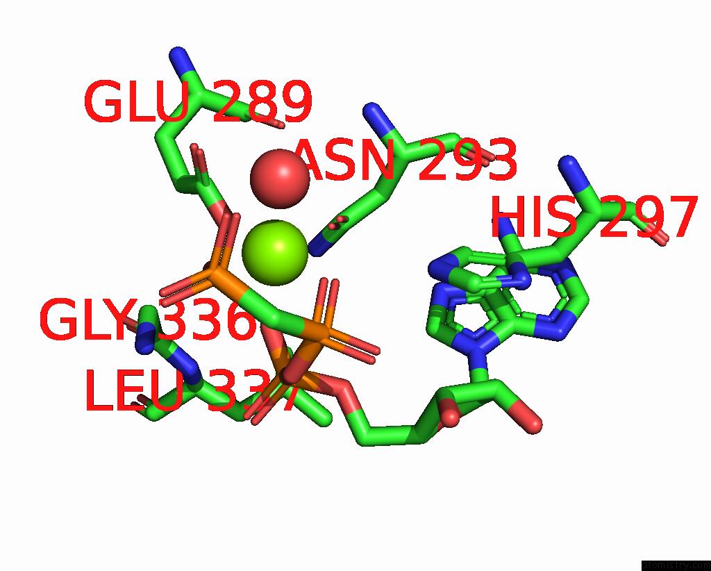
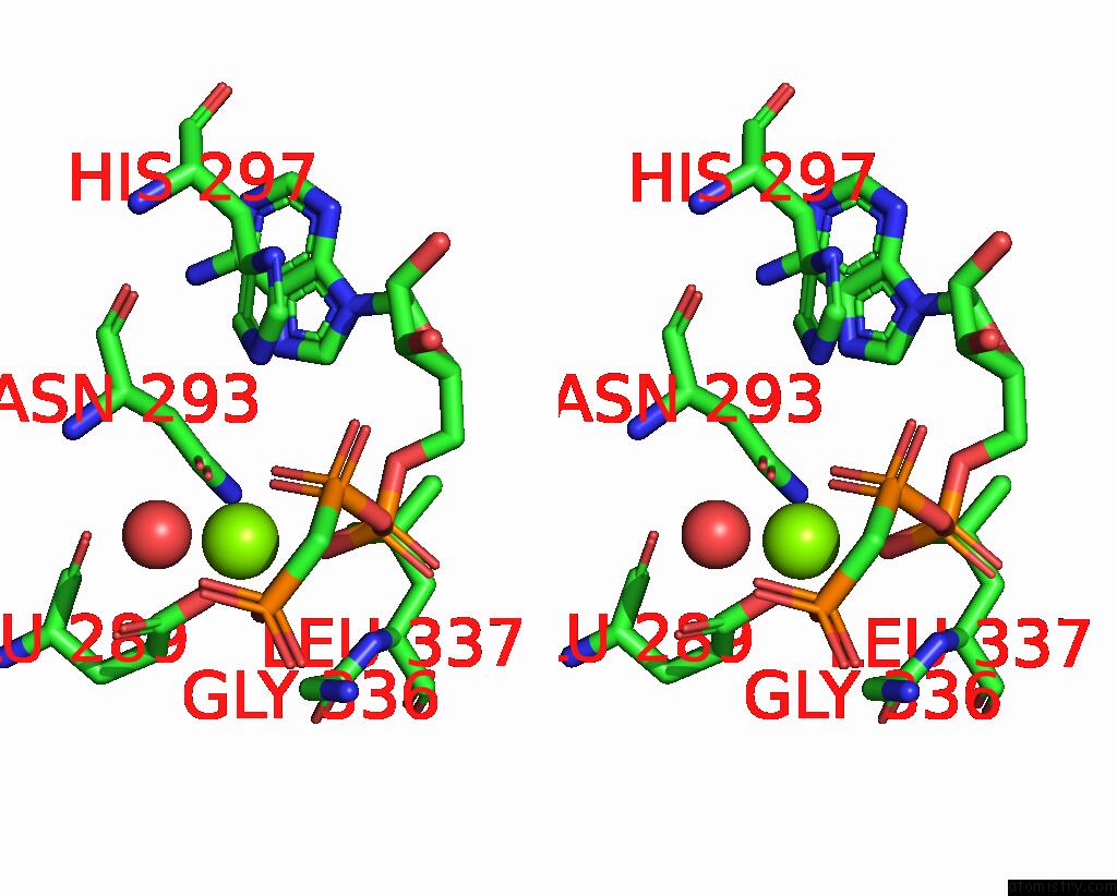
Magnesium binding site 2 out of 4 in 5iuj
Go back to
Magnesium binding site 2 out
of 4 in the Crystal Structure of the Desk-Desr Complex in the Phosphotransfer State with Low MG2+ (20 Mm)
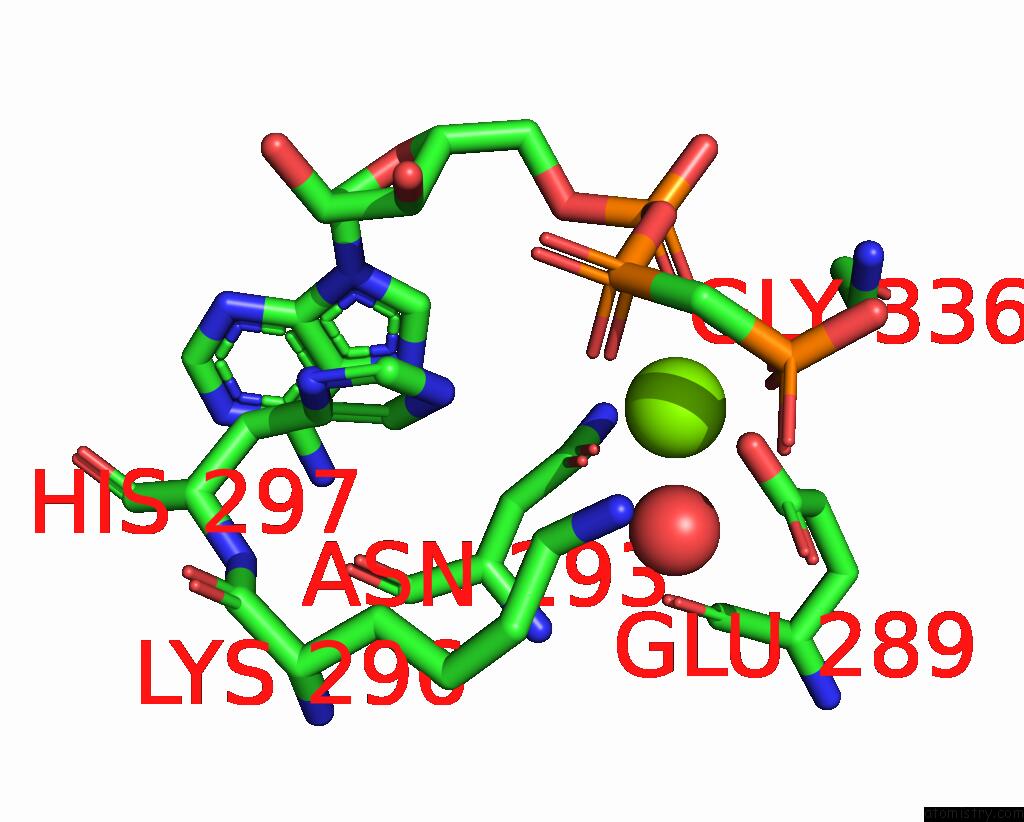
Mono view
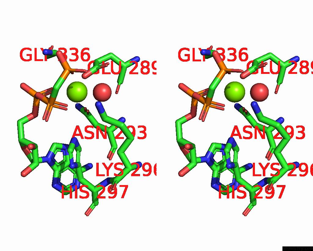
Stereo pair view
Mono view
Stereo pair view
A full contact list of Magnesium with other atoms in the Mg binding
site number 2 of Crystal Structure of the Desk-Desr Complex in the Phosphotransfer State with Low MG2+ (20 Mm) within 5.0Å range:
|
Magnesium binding site 3 out of 4 in 5iuj
Go back to
Magnesium binding site 3 out
of 4 in the Crystal Structure of the Desk-Desr Complex in the Phosphotransfer State with Low MG2+ (20 Mm)
Mono view
Stereo pair view
Mono view
Stereo pair view
A full contact list of Magnesium with other atoms in the Mg binding
site number 3 of Crystal Structure of the Desk-Desr Complex in the Phosphotransfer State with Low MG2+ (20 Mm) within 5.0Å range:
|
Magnesium binding site 4 out of 4 in 5iuj
Go back to
Magnesium binding site 4 out
of 4 in the Crystal Structure of the Desk-Desr Complex in the Phosphotransfer State with Low MG2+ (20 Mm)
Mono view
Stereo pair view
Mono view
Stereo pair view
A full contact list of Magnesium with other atoms in the Mg binding
site number 4 of Crystal Structure of the Desk-Desr Complex in the Phosphotransfer State with Low MG2+ (20 Mm) within 5.0Å range:
|
Reference:
F.Trajtenberg,
J.A.Imelio,
M.R.Machado,
N.Larrieux,
M.A.Marti,
G.Obal,
A.E.Mechaly,
A.Buschiazzo.
Regulation of Signaling Directionality Revealed By 3D Snapshots of A Kinase:Regulator Complex in Action. Elife V. 5 2016.
ISSN: ESSN 2050-084X
PubMed: 27938660
DOI: 10.7554/ELIFE.21422
Page generated: Tue Aug 12 11:37:08 2025
ISSN: ESSN 2050-084X
PubMed: 27938660
DOI: 10.7554/ELIFE.21422
Last articles
Mg in 5MMJMg in 5MRA
Mg in 5MTV
Mg in 5MS0
Mg in 5MRU
Mg in 5MQJ
Mg in 5MQW
Mg in 5MQT
Mg in 5MQL
Mg in 5MQ1