Magnesium »
PDB 5imp-5ivg »
5iuk »
Magnesium in PDB 5iuk: Crystal Structure of the Desk-Desr Complex in the Phosphotransfer State with High MG2+ (150 Mm)
Enzymatic activity of Crystal Structure of the Desk-Desr Complex in the Phosphotransfer State with High MG2+ (150 Mm)
All present enzymatic activity of Crystal Structure of the Desk-Desr Complex in the Phosphotransfer State with High MG2+ (150 Mm):
2.7.13.3;
2.7.13.3;
Protein crystallography data
The structure of Crystal Structure of the Desk-Desr Complex in the Phosphotransfer State with High MG2+ (150 Mm), PDB code: 5iuk
was solved by
F.Trajtenberg,
J.A.Imelio,
N.Larrieux,
A.Buschiazzo,
with X-Ray Crystallography technique. A brief refinement statistics is given in the table below:
| Resolution Low / High (Å) | 39.27 / 2.90 |
| Space group | P 1 21 1 |
| Cell size a, b, c (Å), α, β, γ (°) | 87.930, 115.610, 91.590, 90.00, 116.71, 90.00 |
| R / Rfree (%) | 19.5 / 23.9 |
Other elements in 5iuk:
The structure of Crystal Structure of the Desk-Desr Complex in the Phosphotransfer State with High MG2+ (150 Mm) also contains other interesting chemical elements:
| Potassium | (K) | 2 atoms |
Magnesium Binding Sites:
The binding sites of Magnesium atom in the Crystal Structure of the Desk-Desr Complex in the Phosphotransfer State with High MG2+ (150 Mm)
(pdb code 5iuk). This binding sites where shown within
5.0 Angstroms radius around Magnesium atom.
In total 5 binding sites of Magnesium where determined in the Crystal Structure of the Desk-Desr Complex in the Phosphotransfer State with High MG2+ (150 Mm), PDB code: 5iuk:
Jump to Magnesium binding site number: 1; 2; 3; 4; 5;
In total 5 binding sites of Magnesium where determined in the Crystal Structure of the Desk-Desr Complex in the Phosphotransfer State with High MG2+ (150 Mm), PDB code: 5iuk:
Jump to Magnesium binding site number: 1; 2; 3; 4; 5;
Magnesium binding site 1 out of 5 in 5iuk
Go back to
Magnesium binding site 1 out
of 5 in the Crystal Structure of the Desk-Desr Complex in the Phosphotransfer State with High MG2+ (150 Mm)
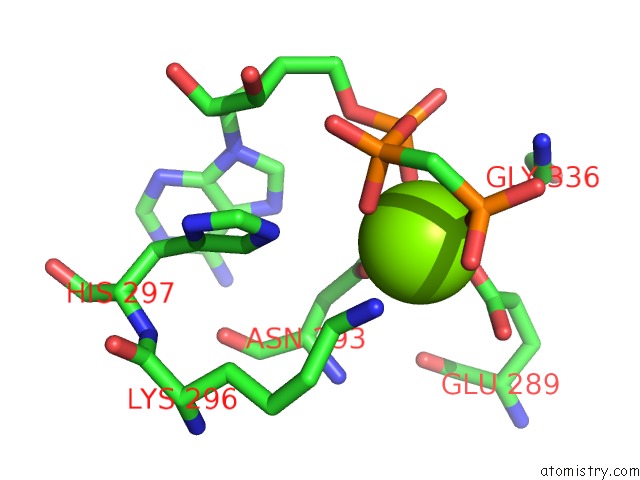
Mono view
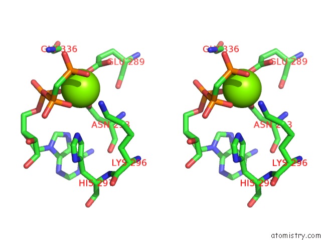
Stereo pair view
Mono view
Stereo pair view
A full contact list of Magnesium with other atoms in the Mg binding
site number 1 of Crystal Structure of the Desk-Desr Complex in the Phosphotransfer State with High MG2+ (150 Mm) within 5.0Å range:
|
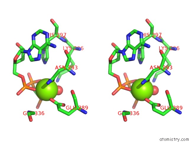
Magnesium binding site 2 out of 5 in 5iuk
Go back to
Magnesium binding site 2 out
of 5 in the Crystal Structure of the Desk-Desr Complex in the Phosphotransfer State with High MG2+ (150 Mm)

Mono view

Stereo pair view
Mono view
Stereo pair view
A full contact list of Magnesium with other atoms in the Mg binding
site number 2 of Crystal Structure of the Desk-Desr Complex in the Phosphotransfer State with High MG2+ (150 Mm) within 5.0Å range:
|
Magnesium binding site 3 out of 5 in 5iuk
Go back to
Magnesium binding site 3 out
of 5 in the Crystal Structure of the Desk-Desr Complex in the Phosphotransfer State with High MG2+ (150 Mm)

Mono view

Stereo pair view
Mono view
Stereo pair view
A full contact list of Magnesium with other atoms in the Mg binding
site number 3 of Crystal Structure of the Desk-Desr Complex in the Phosphotransfer State with High MG2+ (150 Mm) within 5.0Å range:
|
Magnesium binding site 4 out of 5 in 5iuk
Go back to
Magnesium binding site 4 out
of 5 in the Crystal Structure of the Desk-Desr Complex in the Phosphotransfer State with High MG2+ (150 Mm)

Mono view
Stereo pair view
Mono view
Stereo pair view
A full contact list of Magnesium with other atoms in the Mg binding
site number 4 of Crystal Structure of the Desk-Desr Complex in the Phosphotransfer State with High MG2+ (150 Mm) within 5.0Å range:
|
Magnesium binding site 5 out of 5 in 5iuk
Go back to
Magnesium binding site 5 out
of 5 in the Crystal Structure of the Desk-Desr Complex in the Phosphotransfer State with High MG2+ (150 Mm)

Mono view

Stereo pair view
Mono view
Stereo pair view
A full contact list of Magnesium with other atoms in the Mg binding
site number 5 of Crystal Structure of the Desk-Desr Complex in the Phosphotransfer State with High MG2+ (150 Mm) within 5.0Å range:
|
Reference:
F.Trajtenberg,
J.A.Imelio,
M.R.Machado,
N.Larrieux,
M.A.Marti,
G.Obal,
A.E.Mechaly,
A.Buschiazzo.
Regulation of Signaling Directionality Revealed By 3D Snapshots of A Kinase:Regulator Complex in Action. Elife V. 5 2016.
ISSN: ESSN 2050-084X
PubMed: 27938660
DOI: 10.7554/ELIFE.21422
Page generated: Sun Sep 29 16:56:48 2024
ISSN: ESSN 2050-084X
PubMed: 27938660
DOI: 10.7554/ELIFE.21422
Last articles
Zn in 9J0NZn in 9J0O
Zn in 9J0P
Zn in 9FJX
Zn in 9EKB
Zn in 9C0F
Zn in 9CAH
Zn in 9CH0
Zn in 9CH3
Zn in 9CH1