Magnesium in PDB 5jd9: Bacillus Cereus Coth Kinase
Protein crystallography data
The structure of Bacillus Cereus Coth Kinase, PDB code: 5jd9
was solved by
D.R.Tomchick,
V.S.Tagliabracci,
A.Sreelatha,
with X-Ray Crystallography technique. A brief refinement statistics is given in the table below:
Resolution Low / High (Å)
|
26.81 /
1.63
|
Space group
|
P 21 21 21
|
Cell size a, b, c (Å), α, β, γ (°)
|
52.757,
63.516,
118.284,
90.00,
90.00,
90.00
|
R / Rfree (%)
|
15.6 /
17.1
|
Magnesium Binding Sites:
The binding sites of Magnesium atom in the Bacillus Cereus Coth Kinase
(pdb code 5jd9). This binding sites where shown within
5.0 Angstroms radius around Magnesium atom.
In total 4 binding sites of Magnesium where determined in the
Bacillus Cereus Coth Kinase, PDB code: 5jd9:
Jump to Magnesium binding site number:
1;
2;
3;
4;
Magnesium binding site 1 out
of 4 in 5jd9
Go back to
Magnesium Binding Sites List in 5jd9
Magnesium binding site 1 out
of 4 in the Bacillus Cereus Coth Kinase
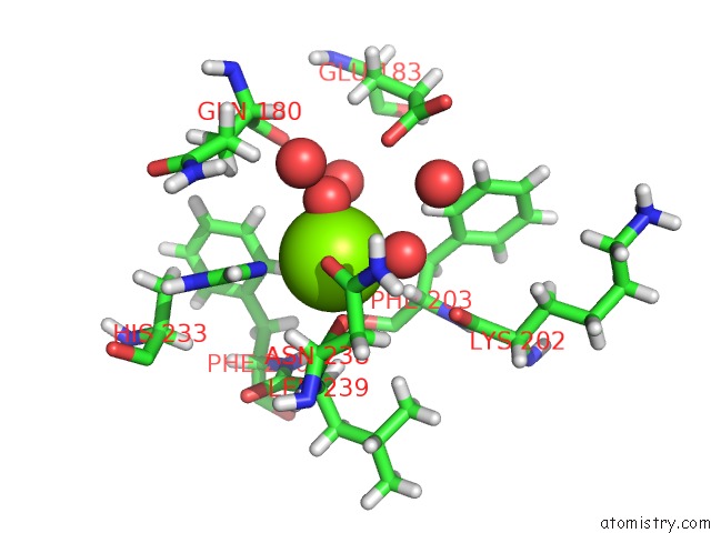
Mono view
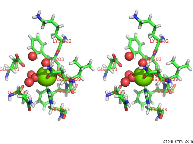
Stereo pair view
|
A full contact list of Magnesium with other atoms in the Mg binding
site number 1 of Bacillus Cereus Coth Kinase within 5.0Å range:
probe
|
atom
|
residue
|
distance (Å)
|
B
|
Occ
|
A:Mg401
b:25.4
occ:1.00
|
O
|
A:HOH526
|
1.9
|
21.6
|
1.0
|
OD1
|
A:ASN238
|
2.0
|
31.5
|
1.0
|
O
|
A:ASN238
|
2.1
|
18.8
|
1.0
|
O
|
A:HOH680
|
2.2
|
23.3
|
1.0
|
O
|
A:HOH652
|
2.3
|
15.4
|
1.0
|
NE2
|
A:HIS233
|
2.4
|
16.9
|
1.0
|
CG
|
A:ASN238
|
2.9
|
36.4
|
1.0
|
C
|
A:ASN238
|
3.0
|
18.6
|
1.0
|
CD2
|
A:HIS233
|
3.2
|
17.5
|
1.0
|
HD2
|
A:HIS233
|
3.3
|
21.0
|
1.0
|
HA
|
A:ASN238
|
3.3
|
20.2
|
1.0
|
CE1
|
A:HIS233
|
3.4
|
19.4
|
1.0
|
CA
|
A:ASN238
|
3.5
|
16.9
|
1.0
|
CB
|
A:ASN238
|
3.5
|
22.9
|
1.0
|
HB3
|
A:ASN238
|
3.5
|
27.4
|
1.0
|
O
|
A:HOH517
|
3.5
|
21.4
|
1.0
|
HE1
|
A:HIS233
|
3.6
|
23.2
|
1.0
|
HA
|
A:PHE203
|
3.9
|
18.5
|
1.0
|
ND2
|
A:ASN238
|
4.0
|
53.2
|
1.0
|
HD21
|
A:ASN238
|
4.1
|
63.9
|
1.0
|
N
|
A:LEU239
|
4.1
|
16.0
|
1.0
|
O
|
A:HOH705
|
4.2
|
28.5
|
1.0
|
HG2
|
A:GLN180
|
4.2
|
24.3
|
1.0
|
HA
|
A:LEU239
|
4.2
|
18.5
|
1.0
|
O
|
A:HOH779
|
4.3
|
38.0
|
1.0
|
OE2
|
A:GLU183
|
4.3
|
54.0
|
1.0
|
HD1
|
A:PHE203
|
4.4
|
24.4
|
1.0
|
HB2
|
A:ASN238
|
4.4
|
27.4
|
1.0
|
CG
|
A:HIS233
|
4.4
|
13.9
|
1.0
|
O
|
A:LYS202
|
4.5
|
19.8
|
1.0
|
ND1
|
A:HIS233
|
4.5
|
19.3
|
1.0
|
HB3
|
A:GLN180
|
4.5
|
20.2
|
1.0
|
HD22
|
A:ASN238
|
4.6
|
63.9
|
1.0
|
CA
|
A:LEU239
|
4.7
|
15.4
|
1.0
|
HD2
|
A:PHE240
|
4.7
|
14.1
|
1.0
|
HD23
|
A:LEU239
|
4.8
|
21.9
|
1.0
|
HE2
|
A:PHE240
|
4.8
|
15.0
|
1.0
|
H
|
A:LEU239
|
4.8
|
19.1
|
1.0
|
O
|
A:PHE203
|
4.8
|
15.2
|
1.0
|
CA
|
A:PHE203
|
4.9
|
15.4
|
1.0
|
N
|
A:ASN238
|
4.9
|
17.5
|
1.0
|
|
Magnesium binding site 2 out
of 4 in 5jd9
Go back to
Magnesium Binding Sites List in 5jd9
Magnesium binding site 2 out
of 4 in the Bacillus Cereus Coth Kinase
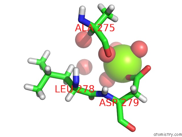
Mono view
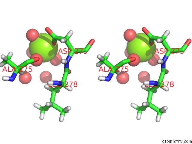
Stereo pair view
|
A full contact list of Magnesium with other atoms in the Mg binding
site number 2 of Bacillus Cereus Coth Kinase within 5.0Å range:
probe
|
atom
|
residue
|
distance (Å)
|
B
|
Occ
|
A:Mg402
b:16.9
occ:1.00
|
OD1
|
A:ASP279
|
2.0
|
21.8
|
1.0
|
O
|
A:HOH589
|
2.1
|
19.9
|
1.0
|
O
|
A:HOH576
|
2.1
|
16.2
|
1.0
|
O
|
A:HOH668
|
2.1
|
20.4
|
1.0
|
O
|
A:HOH822
|
2.1
|
21.9
|
1.0
|
CG
|
A:ASP279
|
3.1
|
25.1
|
1.0
|
OD2
|
A:ASP279
|
3.5
|
21.2
|
1.0
|
O
|
A:HOH802
|
3.8
|
37.7
|
1.0
|
HA
|
A:ASP279
|
4.0
|
18.4
|
1.0
|
O
|
A:HOH834
|
4.1
|
18.1
|
1.0
|
O
|
A:ALA275
|
4.2
|
14.0
|
1.0
|
O
|
A:LEU278
|
4.3
|
21.4
|
1.0
|
CB
|
A:ASP279
|
4.3
|
17.5
|
1.0
|
CA
|
A:ASP279
|
4.5
|
15.3
|
1.0
|
N
|
A:ASP279
|
4.6
|
14.7
|
1.0
|
C
|
A:LEU278
|
4.7
|
14.9
|
1.0
|
HB1
|
A:ALA275
|
4.7
|
16.2
|
1.0
|
HA
|
A:ALA275
|
4.8
|
14.4
|
1.0
|
HB2
|
A:ASP279
|
4.9
|
21.1
|
1.0
|
HB3
|
A:ASP279
|
4.9
|
21.1
|
1.0
|
H
|
A:ASP279
|
5.0
|
17.6
|
1.0
|
|
Magnesium binding site 3 out
of 4 in 5jd9
Go back to
Magnesium Binding Sites List in 5jd9
Magnesium binding site 3 out
of 4 in the Bacillus Cereus Coth Kinase
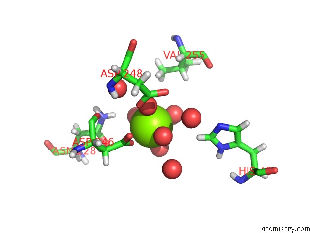
Mono view
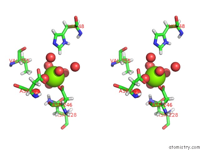
Stereo pair view
|
A full contact list of Magnesium with other atoms in the Mg binding
site number 3 of Bacillus Cereus Coth Kinase within 5.0Å range:
probe
|
atom
|
residue
|
distance (Å)
|
B
|
Occ
|
A:Mg403
b:28.9
occ:1.00
|
O
|
A:HOH674
|
2.1
|
40.1
|
1.0
|
O
|
A:HOH506
|
2.1
|
31.3
|
1.0
|
OD2
|
A:ASP246
|
2.2
|
32.9
|
1.0
|
O
|
A:HOH555
|
2.2
|
26.9
|
1.0
|
O
|
A:HOH825
|
2.2
|
28.2
|
1.0
|
O
|
A:HOH717
|
2.3
|
30.2
|
1.0
|
CG
|
A:ASP246
|
3.0
|
26.3
|
1.0
|
OD1
|
A:ASP246
|
3.2
|
40.0
|
1.0
|
O
|
A:HOH827
|
3.8
|
41.2
|
1.0
|
HD21
|
A:ASN228
|
3.9
|
29.8
|
1.0
|
HE1
|
A:HIS48
|
3.9
|
25.5
|
1.0
|
O
|
A:HOH586
|
4.1
|
15.4
|
1.0
|
OD1
|
A:ASP248
|
4.2
|
21.1
|
1.0
|
HG12
|
A:VAL255
|
4.3
|
22.5
|
1.0
|
OD1
|
A:ASN228
|
4.4
|
17.3
|
1.0
|
CB
|
A:ASP246
|
4.4
|
8.9
|
1.0
|
O
|
A:HOH819
|
4.5
|
28.8
|
1.0
|
HB3
|
A:ASP246
|
4.5
|
10.7
|
1.0
|
O
|
A:ASP246
|
4.5
|
11.0
|
1.0
|
O
|
A:HOH691
|
4.6
|
36.7
|
1.0
|
ND2
|
A:ASN228
|
4.6
|
24.8
|
1.0
|
CE1
|
A:HIS48
|
4.8
|
21.2
|
1.0
|
HG11
|
A:VAL255
|
4.9
|
22.5
|
1.0
|
HB2
|
A:ASP246
|
4.9
|
10.7
|
1.0
|
CG
|
A:ASN228
|
4.9
|
16.8
|
1.0
|
|
Magnesium binding site 4 out
of 4 in 5jd9
Go back to
Magnesium Binding Sites List in 5jd9
Magnesium binding site 4 out
of 4 in the Bacillus Cereus Coth Kinase
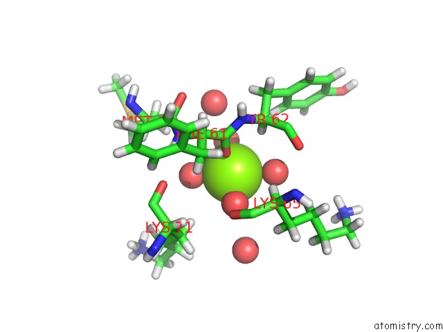
Mono view
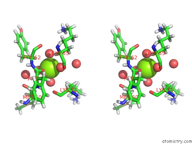
Stereo pair view
|
A full contact list of Magnesium with other atoms in the Mg binding
site number 4 of Bacillus Cereus Coth Kinase within 5.0Å range:
probe
|
atom
|
residue
|
distance (Å)
|
B
|
Occ
|
A:Mg404
b:45.0
occ:1.00
|
O
|
A:PHE61
|
2.0
|
14.4
|
1.0
|
O
|
A:HOH547
|
2.0
|
21.6
|
1.0
|
O
|
A:HOH650
|
2.3
|
25.2
|
1.0
|
O
|
A:HOH760
|
2.3
|
32.8
|
1.0
|
O
|
A:HOH629
|
2.4
|
31.4
|
1.0
|
HA
|
A:TYR62
|
3.1
|
16.0
|
1.0
|
C
|
A:PHE61
|
3.2
|
11.8
|
1.0
|
HB2
|
A:LYS71
|
3.8
|
20.4
|
1.0
|
CA
|
A:TYR62
|
3.9
|
13.4
|
1.0
|
N
|
A:TYR62
|
4.0
|
12.1
|
1.0
|
HA
|
A:LYS65
|
4.1
|
19.1
|
1.0
|
O
|
A:HOH538
|
4.2
|
24.2
|
1.0
|
O
|
A:LYS65
|
4.3
|
16.9
|
1.0
|
CA
|
A:PHE61
|
4.3
|
10.9
|
1.0
|
HD1
|
A:TYR62
|
4.4
|
20.7
|
1.0
|
H
|
A:PHE61
|
4.4
|
12.5
|
1.0
|
N
|
A:PHE61
|
4.4
|
10.4
|
1.0
|
O
|
A:LYS71
|
4.4
|
11.8
|
1.0
|
O
|
A:HOH832
|
4.4
|
44.5
|
1.0
|
HB2
|
A:PHE61
|
4.5
|
13.2
|
1.0
|
O
|
A:TYR62
|
4.5
|
16.3
|
1.0
|
HB3
|
A:MET60
|
4.5
|
17.1
|
1.0
|
HB3
|
A:LYS65
|
4.7
|
26.2
|
1.0
|
C
|
A:TYR62
|
4.7
|
14.3
|
1.0
|
CB
|
A:LYS71
|
4.7
|
17.0
|
1.0
|
HG2
|
A:LYS65
|
4.7
|
33.2
|
1.0
|
HD2
|
A:LYS71
|
4.7
|
36.9
|
1.0
|
H
|
A:TYR62
|
4.8
|
14.6
|
1.0
|
CA
|
A:LYS65
|
4.9
|
15.9
|
1.0
|
HB2
|
A:TYR62
|
4.9
|
16.0
|
1.0
|
CB
|
A:PHE61
|
4.9
|
11.0
|
1.0
|
C
|
A:MET60
|
4.9
|
12.1
|
1.0
|
CB
|
A:TYR62
|
5.0
|
13.3
|
1.0
|
|
Reference:
K.B.Nguyen,
A.Sreelatha,
E.S.Durrant,
J.Lopez-Garrido,
A.Muszewska,
M.Dudkiewicz,
M.Grynberg,
S.Yee,
K.Pogliano,
D.R.Tomchick,
K.Pawowski,
J.E.Dixon,
V.S.Tagliabracci.
Phosphorylation of Spore Coat Proteins By A Family of Atypical Protein Kinases. Proc.Natl.Acad.Sci.Usa V. 113 E3482 2016.
ISSN: ESSN 1091-6490
PubMed: 27185916
DOI: 10.1073/PNAS.1605917113
Page generated: Sun Sep 29 17:43:23 2024
|