Magnesium »
PDB 5jca-5jo2 »
5jmv »
Magnesium in PDB 5jmv: Crystal Structure of MJKAE1-PFUPCC1 Complex
Enzymatic activity of Crystal Structure of MJKAE1-PFUPCC1 Complex
Protein crystallography data
The structure of Crystal Structure of MJKAE1-PFUPCC1 Complex, PDB code: 5jmv
was solved by
L.Wan,
F.Sicheri,
with X-Ray Crystallography technique. A brief refinement statistics is given in the table below:
| Resolution Low / High (Å) | 48.24 / 3.39 |
| Space group | P 43 21 2 |
| Cell size a, b, c (Å), α, β, γ (°) | 121.913, 121.913, 310.613, 90.00, 90.00, 90.00 |
| R / Rfree (%) | 20 / 24.3 |
Magnesium Binding Sites:
The binding sites of Magnesium atom in the Crystal Structure of MJKAE1-PFUPCC1 Complex
(pdb code 5jmv). This binding sites where shown within
5.0 Angstroms radius around Magnesium atom.
In total 3 binding sites of Magnesium where determined in the Crystal Structure of MJKAE1-PFUPCC1 Complex, PDB code: 5jmv:
Jump to Magnesium binding site number: 1; 2; 3;
In total 3 binding sites of Magnesium where determined in the Crystal Structure of MJKAE1-PFUPCC1 Complex, PDB code: 5jmv:
Jump to Magnesium binding site number: 1; 2; 3;
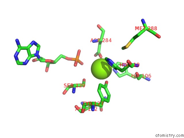
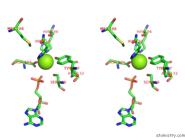
Magnesium binding site 1 out of 3 in 5jmv
Go back to
Magnesium binding site 1 out
of 3 in the Crystal Structure of MJKAE1-PFUPCC1 Complex
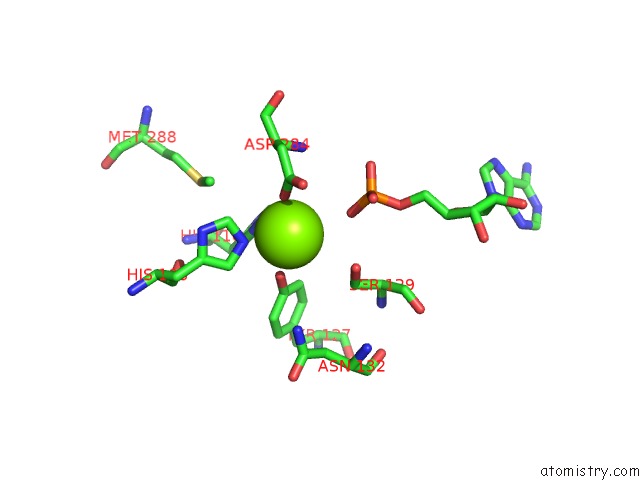
Mono view
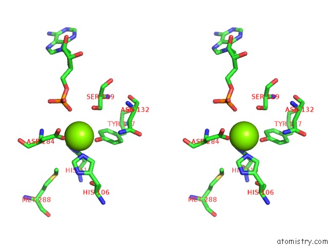
Stereo pair view
Mono view
Stereo pair view
A full contact list of Magnesium with other atoms in the Mg binding
site number 1 of Crystal Structure of MJKAE1-PFUPCC1 Complex within 5.0Å range:
|
Magnesium binding site 2 out of 3 in 5jmv
Go back to
Magnesium binding site 2 out
of 3 in the Crystal Structure of MJKAE1-PFUPCC1 Complex
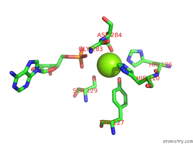
Mono view
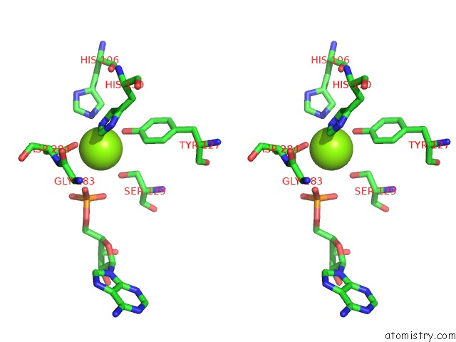
Stereo pair view
Mono view
Stereo pair view
A full contact list of Magnesium with other atoms in the Mg binding
site number 2 of Crystal Structure of MJKAE1-PFUPCC1 Complex within 5.0Å range:
|
Magnesium binding site 3 out of 3 in 5jmv
Go back to
Magnesium binding site 3 out
of 3 in the Crystal Structure of MJKAE1-PFUPCC1 Complex
Mono view
Stereo pair view
Mono view
Stereo pair view
A full contact list of Magnesium with other atoms in the Mg binding
site number 3 of Crystal Structure of MJKAE1-PFUPCC1 Complex within 5.0Å range:
|
Reference:
L.C.Wan,
M.C.Pillon,
N.Thevakumaran,
Y.Sun,
A.Chakrabartty,
A.Guarne,
I.Kurinov,
D.Durocher,
F.Sicheri.
Structural and Functional Characterization of Keops Dimerization By PCC1 and Its Role in T6A Biosynthesis. Nucleic Acids Res. V. 44 6971 2016.
ISSN: ESSN 1362-4962
PubMed: 27302132
DOI: 10.1093/NAR/GKW542
Page generated: Sun Sep 29 17:52:50 2024
ISSN: ESSN 1362-4962
PubMed: 27302132
DOI: 10.1093/NAR/GKW542
Last articles
Zn in 9J0NZn in 9J0O
Zn in 9J0P
Zn in 9FJX
Zn in 9EKB
Zn in 9C0F
Zn in 9CAH
Zn in 9CH0
Zn in 9CH3
Zn in 9CH1