Magnesium »
PDB 5jp0-5k0l »
5jye »
Magnesium in PDB 5jye: Structures of Streptococcus Agalactiae Gbs Gapdh in Different Enzymatic States
Protein crystallography data
The structure of Structures of Streptococcus Agalactiae Gbs Gapdh in Different Enzymatic States, PDB code: 5jye
was solved by
N.Schormann,
D.Chattopadhyay,
with X-Ray Crystallography technique. A brief refinement statistics is given in the table below:
| Resolution Low / High (Å) | 80.91 / 2.23 |
| Space group | P 1 21 1 |
| Cell size a, b, c (Å), α, β, γ (°) | 78.247, 107.358, 87.904, 90.00, 113.01, 90.00 |
| R / Rfree (%) | 20.7 / 24.9 |
Magnesium Binding Sites:
The binding sites of Magnesium atom in the Structures of Streptococcus Agalactiae Gbs Gapdh in Different Enzymatic States
(pdb code 5jye). This binding sites where shown within
5.0 Angstroms radius around Magnesium atom.
In total 2 binding sites of Magnesium where determined in the Structures of Streptococcus Agalactiae Gbs Gapdh in Different Enzymatic States, PDB code: 5jye:
Jump to Magnesium binding site number: 1; 2;
In total 2 binding sites of Magnesium where determined in the Structures of Streptococcus Agalactiae Gbs Gapdh in Different Enzymatic States, PDB code: 5jye:
Jump to Magnesium binding site number: 1; 2;
Magnesium binding site 1 out of 2 in 5jye
Go back to
Magnesium binding site 1 out
of 2 in the Structures of Streptococcus Agalactiae Gbs Gapdh in Different Enzymatic States
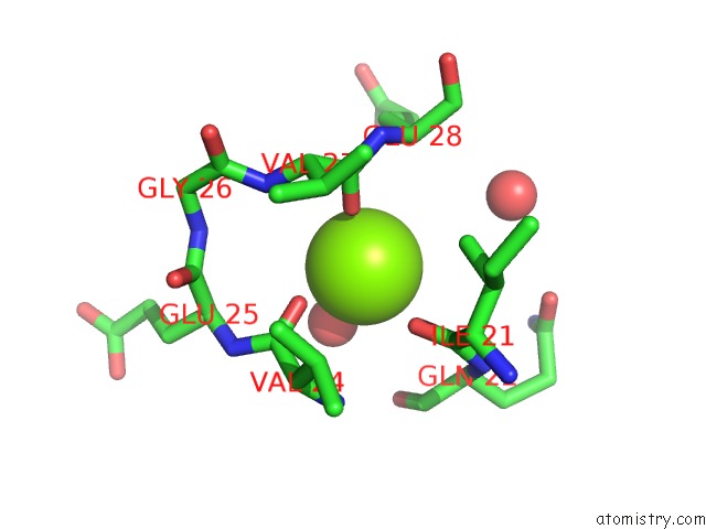
Mono view
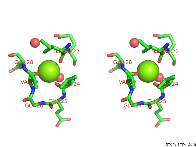
Stereo pair view
Mono view
Stereo pair view
A full contact list of Magnesium with other atoms in the Mg binding
site number 1 of Structures of Streptococcus Agalactiae Gbs Gapdh in Different Enzymatic States within 5.0Å range:
|
Magnesium binding site 2 out of 2 in 5jye
Go back to
Magnesium binding site 2 out
of 2 in the Structures of Streptococcus Agalactiae Gbs Gapdh in Different Enzymatic States
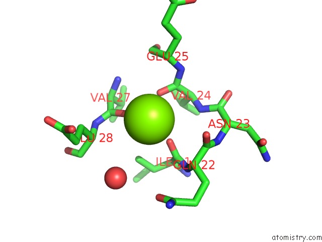
Mono view
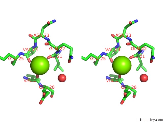
Stereo pair view
Mono view
Stereo pair view
A full contact list of Magnesium with other atoms in the Mg binding
site number 2 of Structures of Streptococcus Agalactiae Gbs Gapdh in Different Enzymatic States within 5.0Å range:
|
Reference:
N.Schormann,
C.A.Ayres,
A.Fry,
T.J.Green,
S.Banerjee,
G.C.Ulett,
D.Chattopadhyay.
Crystal Structures of Group B Streptococcus Glyceraldehyde-3-Phosphate Dehydrogenase: Apo-Form, Binary and Ternary Complexes. Plos One V. 11 65917 2016.
ISSN: ESSN 1932-6203
PubMed: 27875551
DOI: 10.1371/JOURNAL.PONE.0165917
Page generated: Sun Sep 29 18:01:54 2024
ISSN: ESSN 1932-6203
PubMed: 27875551
DOI: 10.1371/JOURNAL.PONE.0165917
Last articles
Zn in 9J0NZn in 9J0O
Zn in 9J0P
Zn in 9FJX
Zn in 9EKB
Zn in 9C0F
Zn in 9CAH
Zn in 9CH0
Zn in 9CH3
Zn in 9CH1