Magnesium »
PDB 5k1i-5kfu »
5kdl »
Magnesium in PDB 5kdl: Crystal Structure of the 4 Alanine Insertion Variant of the Gi ALPHA1 Subunit Bound to Gtpgammas
Protein crystallography data
The structure of Crystal Structure of the 4 Alanine Insertion Variant of the Gi ALPHA1 Subunit Bound to Gtpgammas, PDB code: 5kdl
was solved by
A.I.Kaya,
A.D.Lokits,
J.Gilbert,
T.M.Iverson,
J.Meiler,
H.E.Hamm,
with X-Ray Crystallography technique. A brief refinement statistics is given in the table below:
| Resolution Low / High (Å) | 47.82 / 2.67 |
| Space group | P 1 21 1 |
| Cell size a, b, c (Å), α, β, γ (°) | 61.742, 77.351, 73.095, 90.00, 99.86, 90.00 |
| R / Rfree (%) | 20.9 / 26.5 |
Magnesium Binding Sites:
The binding sites of Magnesium atom in the Crystal Structure of the 4 Alanine Insertion Variant of the Gi ALPHA1 Subunit Bound to Gtpgammas
(pdb code 5kdl). This binding sites where shown within
5.0 Angstroms radius around Magnesium atom.
In total 2 binding sites of Magnesium where determined in the Crystal Structure of the 4 Alanine Insertion Variant of the Gi ALPHA1 Subunit Bound to Gtpgammas, PDB code: 5kdl:
Jump to Magnesium binding site number: 1; 2;
In total 2 binding sites of Magnesium where determined in the Crystal Structure of the 4 Alanine Insertion Variant of the Gi ALPHA1 Subunit Bound to Gtpgammas, PDB code: 5kdl:
Jump to Magnesium binding site number: 1; 2;
Magnesium binding site 1 out of 2 in 5kdl
Go back to
Magnesium binding site 1 out
of 2 in the Crystal Structure of the 4 Alanine Insertion Variant of the Gi ALPHA1 Subunit Bound to Gtpgammas
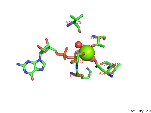
Mono view
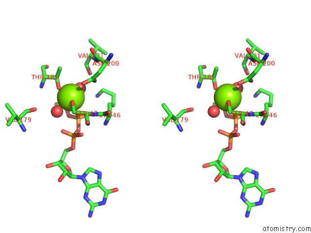
Stereo pair view
Mono view
Stereo pair view
A full contact list of Magnesium with other atoms in the Mg binding
site number 1 of Crystal Structure of the 4 Alanine Insertion Variant of the Gi ALPHA1 Subunit Bound to Gtpgammas within 5.0Å range:
|
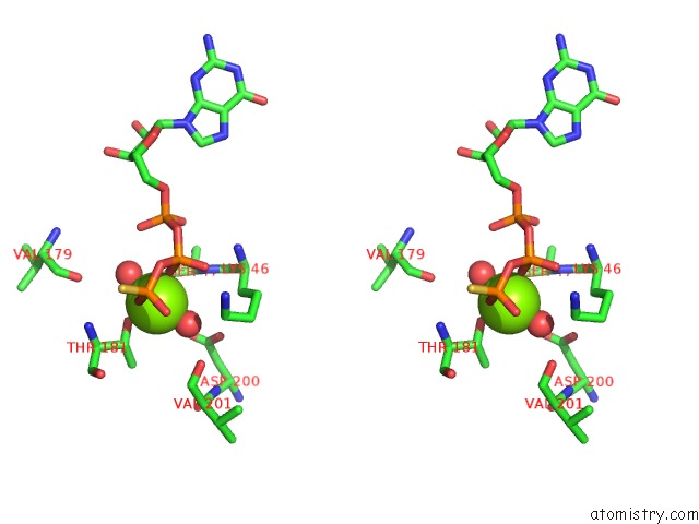
Magnesium binding site 2 out of 2 in 5kdl
Go back to
Magnesium binding site 2 out
of 2 in the Crystal Structure of the 4 Alanine Insertion Variant of the Gi ALPHA1 Subunit Bound to Gtpgammas
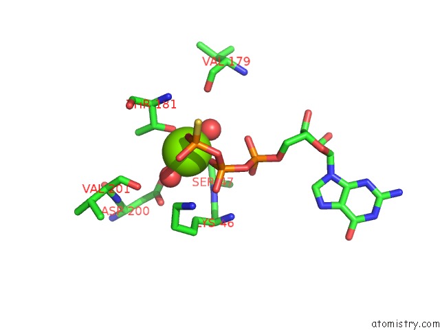
Mono view
Stereo pair view
Mono view
Stereo pair view
A full contact list of Magnesium with other atoms in the Mg binding
site number 2 of Crystal Structure of the 4 Alanine Insertion Variant of the Gi ALPHA1 Subunit Bound to Gtpgammas within 5.0Å range:
|
Reference:
A.I.Kaya,
A.D.Lokits,
J.A.Gilbert,
T.M.Iverson,
J.Meiler,
H.E.Hamm.
A Conserved Hydrophobic Core in G Alpha I1 Regulates G Protein Activation and Release From Activated Receptor. J.Biol.Chem. V. 291 19674 2016.
ISSN: ESSN 1083-351X
PubMed: 27462082
DOI: 10.1074/JBC.M116.745513
Page generated: Tue Aug 12 13:05:42 2025
ISSN: ESSN 1083-351X
PubMed: 27462082
DOI: 10.1074/JBC.M116.745513
Last articles
Mg in 6ZOLMg in 6ZND
Mg in 6ZNW
Mg in 6ZJB
Mg in 6ZN7
Mg in 6ZN4
Mg in 6ZMD
Mg in 6ZLI
Mg in 6ZM2
Mg in 6ZL7