Magnesium »
PDB 5kfu-5ktd »
5knx »
Magnesium in PDB 5knx: Crystal Structure of E. Coli Hypoxanthine Phosphoribosyltransferase in Complexed with {[(2-[(Hypoxanthin-9H-Yl)Methyl]Propane-1,3-Diyl) Bis(Oxy)]Bis- (Methylene)}Diphosphonic Acid
Protein crystallography data
The structure of Crystal Structure of E. Coli Hypoxanthine Phosphoribosyltransferase in Complexed with {[(2-[(Hypoxanthin-9H-Yl)Methyl]Propane-1,3-Diyl) Bis(Oxy)]Bis- (Methylene)}Diphosphonic Acid, PDB code: 5knx
was solved by
W.S.Eng,
D.T.Keough,
D.Hockova,
Z.Janeba,
L.W.Guddat,
with X-Ray Crystallography technique. A brief refinement statistics is given in the table below:
| Resolution Low / High (Å) | 55.83 / 2.40 |
| Space group | P 31 2 1 |
| Cell size a, b, c (Å), α, β, γ (°) | 85.380, 85.380, 167.475, 90.00, 90.00, 120.00 |
| R / Rfree (%) | 18.2 / 22.1 |
Magnesium Binding Sites:
The binding sites of Magnesium atom in the Crystal Structure of E. Coli Hypoxanthine Phosphoribosyltransferase in Complexed with {[(2-[(Hypoxanthin-9H-Yl)Methyl]Propane-1,3-Diyl) Bis(Oxy)]Bis- (Methylene)}Diphosphonic Acid
(pdb code 5knx). This binding sites where shown within
5.0 Angstroms radius around Magnesium atom.
In total 3 binding sites of Magnesium where determined in the Crystal Structure of E. Coli Hypoxanthine Phosphoribosyltransferase in Complexed with {[(2-[(Hypoxanthin-9H-Yl)Methyl]Propane-1,3-Diyl) Bis(Oxy)]Bis- (Methylene)}Diphosphonic Acid, PDB code: 5knx:
Jump to Magnesium binding site number: 1; 2; 3;
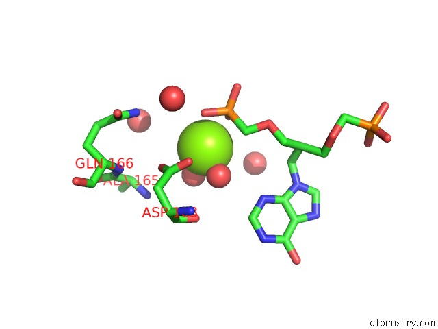
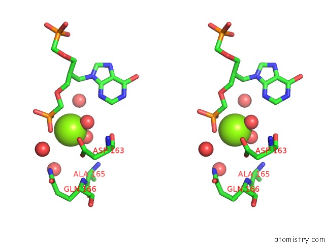
Magnesium binding site 1 out of 3 in 5knx
Go back to
Magnesium binding site 1 out
of 3 in the Crystal Structure of E. Coli Hypoxanthine Phosphoribosyltransferase in Complexed with {[(2-[(Hypoxanthin-9H-Yl)Methyl]Propane-1,3-Diyl) Bis(Oxy)]Bis- (Methylene)}Diphosphonic Acid
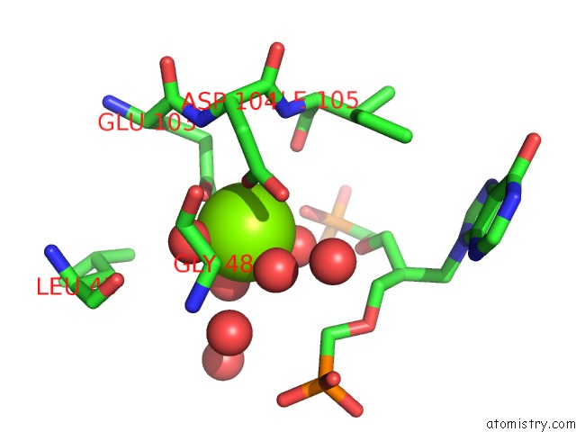
Mono view
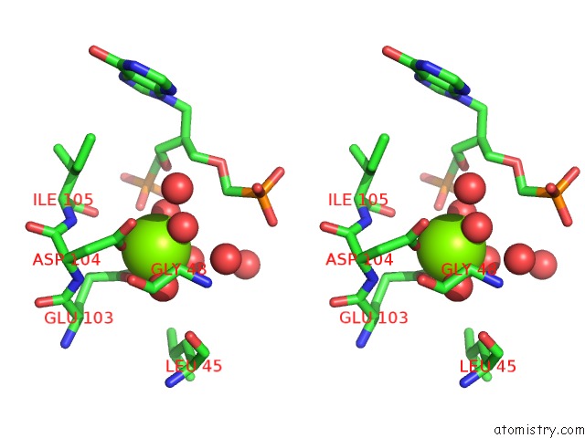
Stereo pair view
Mono view
Stereo pair view
A full contact list of Magnesium with other atoms in the Mg binding
site number 1 of Crystal Structure of E. Coli Hypoxanthine Phosphoribosyltransferase in Complexed with {[(2-[(Hypoxanthin-9H-Yl)Methyl]Propane-1,3-Diyl) Bis(Oxy)]Bis- (Methylene)}Diphosphonic Acid within 5.0Å range:
|
Magnesium binding site 2 out of 3 in 5knx
Go back to
Magnesium binding site 2 out
of 3 in the Crystal Structure of E. Coli Hypoxanthine Phosphoribosyltransferase in Complexed with {[(2-[(Hypoxanthin-9H-Yl)Methyl]Propane-1,3-Diyl) Bis(Oxy)]Bis- (Methylene)}Diphosphonic Acid
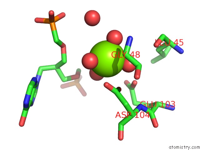
Mono view
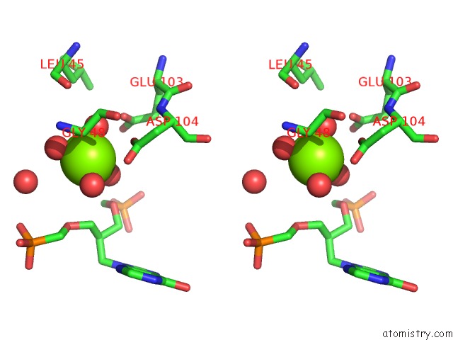
Stereo pair view
Mono view
Stereo pair view
A full contact list of Magnesium with other atoms in the Mg binding
site number 2 of Crystal Structure of E. Coli Hypoxanthine Phosphoribosyltransferase in Complexed with {[(2-[(Hypoxanthin-9H-Yl)Methyl]Propane-1,3-Diyl) Bis(Oxy)]Bis- (Methylene)}Diphosphonic Acid within 5.0Å range:
|
Magnesium binding site 3 out of 3 in 5knx
Go back to
Magnesium binding site 3 out
of 3 in the Crystal Structure of E. Coli Hypoxanthine Phosphoribosyltransferase in Complexed with {[(2-[(Hypoxanthin-9H-Yl)Methyl]Propane-1,3-Diyl) Bis(Oxy)]Bis- (Methylene)}Diphosphonic Acid
Mono view
Stereo pair view
Mono view
Stereo pair view
A full contact list of Magnesium with other atoms in the Mg binding
site number 3 of Crystal Structure of E. Coli Hypoxanthine Phosphoribosyltransferase in Complexed with {[(2-[(Hypoxanthin-9H-Yl)Methyl]Propane-1,3-Diyl) Bis(Oxy)]Bis- (Methylene)}Diphosphonic Acid within 5.0Å range:
|
Reference:
W.S.Eng,
D.Hockova,
P.Spacek,
O.Baszczynski,
Z.Janeba,
L.Naesens,
D.T.Keough,
L.W.Guddat.
Crystal Structures of Acyclic Nucleoside Phosphonates in Complex with Escherichia Coli Hypoxanthine Phosphoribosyltransferase Chemistryselect V. 1 6267 2016.
ISSN: ESSN 2365-6549
DOI: 10.1002/SLCT.201601679
Page generated: Sun Sep 29 19:10:11 2024
ISSN: ESSN 2365-6549
DOI: 10.1002/SLCT.201601679
Last articles
Zn in 9J0NZn in 9J0O
Zn in 9J0P
Zn in 9FJX
Zn in 9EKB
Zn in 9C0F
Zn in 9CAH
Zn in 9CH0
Zn in 9CH3
Zn in 9CH1