Magnesium »
PDB 5kfv-5ktj »
5kny »
Magnesium in PDB 5kny: Crystal Structure of Mycobacterium Tuberculosis Hypoxanthine Guanine Phosphoribosyltransferase in Complex with (3-((3R,4R)-3-(Guanin-9- Yl)-4-((S)-2-Hydroxy-2-Phosphonoethoxy)Pyrrolidin-1-Yl)-3-Oxopropyl) Phosphonic Acid
Enzymatic activity of Crystal Structure of Mycobacterium Tuberculosis Hypoxanthine Guanine Phosphoribosyltransferase in Complex with (3-((3R,4R)-3-(Guanin-9- Yl)-4-((S)-2-Hydroxy-2-Phosphonoethoxy)Pyrrolidin-1-Yl)-3-Oxopropyl) Phosphonic Acid
All present enzymatic activity of Crystal Structure of Mycobacterium Tuberculosis Hypoxanthine Guanine Phosphoribosyltransferase in Complex with (3-((3R,4R)-3-(Guanin-9- Yl)-4-((S)-2-Hydroxy-2-Phosphonoethoxy)Pyrrolidin-1-Yl)-3-Oxopropyl) Phosphonic Acid:
2.4.2.8;
2.4.2.8;
Protein crystallography data
The structure of Crystal Structure of Mycobacterium Tuberculosis Hypoxanthine Guanine Phosphoribosyltransferase in Complex with (3-((3R,4R)-3-(Guanin-9- Yl)-4-((S)-2-Hydroxy-2-Phosphonoethoxy)Pyrrolidin-1-Yl)-3-Oxopropyl) Phosphonic Acid, PDB code: 5kny
was solved by
W.S.Eng,
D.Rejman,
D.T.Keough,
L.W.Guddat,
with X-Ray Crystallography technique. A brief refinement statistics is given in the table below:
| Resolution Low / High (Å) | 44.77 / 2.91 |
| Space group | P 1 21 1 |
| Cell size a, b, c (Å), α, β, γ (°) | 54.523, 86.075, 79.824, 90.00, 105.95, 90.00 |
| R / Rfree (%) | 20.1 / 26.2 |
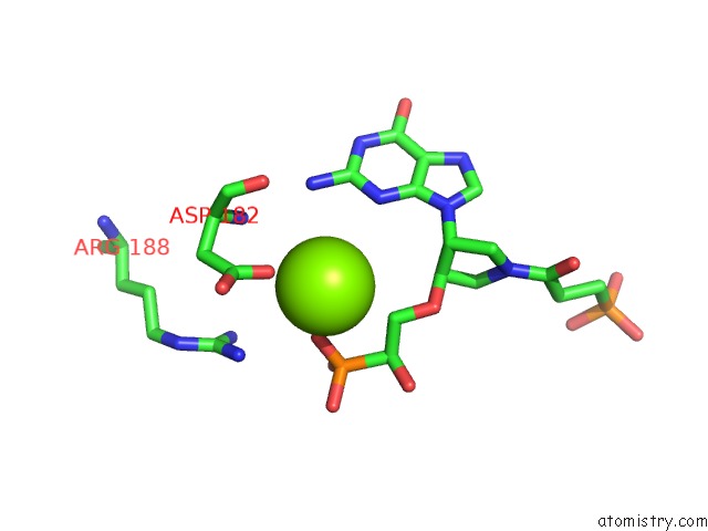
Magnesium Binding Sites:
The binding sites of Magnesium atom in the Crystal Structure of Mycobacterium Tuberculosis Hypoxanthine Guanine Phosphoribosyltransferase in Complex with (3-((3R,4R)-3-(Guanin-9- Yl)-4-((S)-2-Hydroxy-2-Phosphonoethoxy)Pyrrolidin-1-Yl)-3-Oxopropyl) Phosphonic Acid
(pdb code 5kny). This binding sites where shown within
5.0 Angstroms radius around Magnesium atom.
In total 4 binding sites of Magnesium where determined in the Crystal Structure of Mycobacterium Tuberculosis Hypoxanthine Guanine Phosphoribosyltransferase in Complex with (3-((3R,4R)-3-(Guanin-9- Yl)-4-((S)-2-Hydroxy-2-Phosphonoethoxy)Pyrrolidin-1-Yl)-3-Oxopropyl) Phosphonic Acid, PDB code: 5kny:
Jump to Magnesium binding site number: 1; 2; 3; 4;
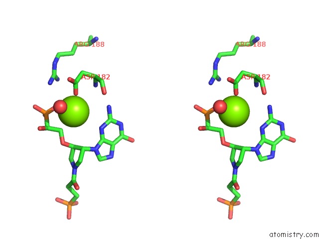
Magnesium binding site 1 out of 4 in 5kny
Go back to
Magnesium binding site 1 out
of 4 in the Crystal Structure of Mycobacterium Tuberculosis Hypoxanthine Guanine Phosphoribosyltransferase in Complex with (3-((3R,4R)-3-(Guanin-9- Yl)-4-((S)-2-Hydroxy-2-Phosphonoethoxy)Pyrrolidin-1-Yl)-3-Oxopropyl) Phosphonic Acid
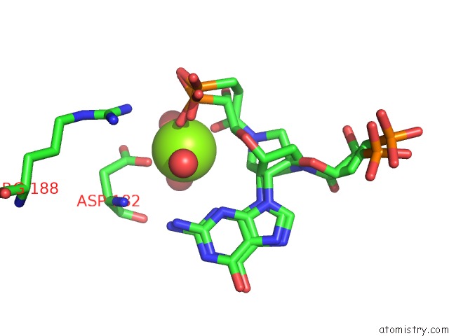
Mono view
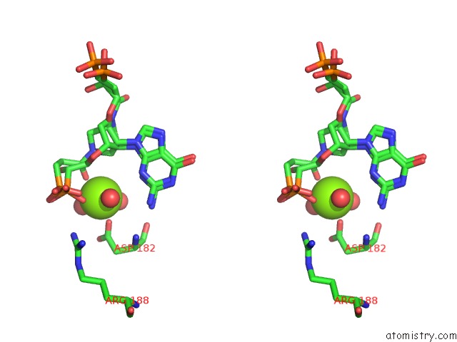
Stereo pair view
Mono view
Stereo pair view
A full contact list of Magnesium with other atoms in the Mg binding
site number 1 of Crystal Structure of Mycobacterium Tuberculosis Hypoxanthine Guanine Phosphoribosyltransferase in Complex with (3-((3R,4R)-3-(Guanin-9- Yl)-4-((S)-2-Hydroxy-2-Phosphonoethoxy)Pyrrolidin-1-Yl)-3-Oxopropyl) Phosphonic Acid within 5.0Å range:
|
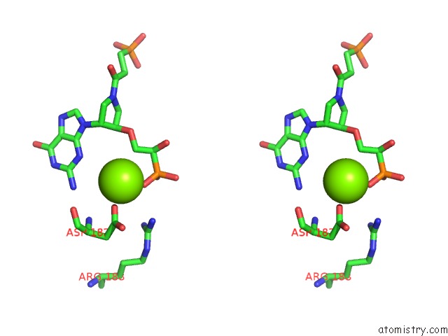
Magnesium binding site 2 out of 4 in 5kny
Go back to
Magnesium binding site 2 out
of 4 in the Crystal Structure of Mycobacterium Tuberculosis Hypoxanthine Guanine Phosphoribosyltransferase in Complex with (3-((3R,4R)-3-(Guanin-9- Yl)-4-((S)-2-Hydroxy-2-Phosphonoethoxy)Pyrrolidin-1-Yl)-3-Oxopropyl) Phosphonic Acid
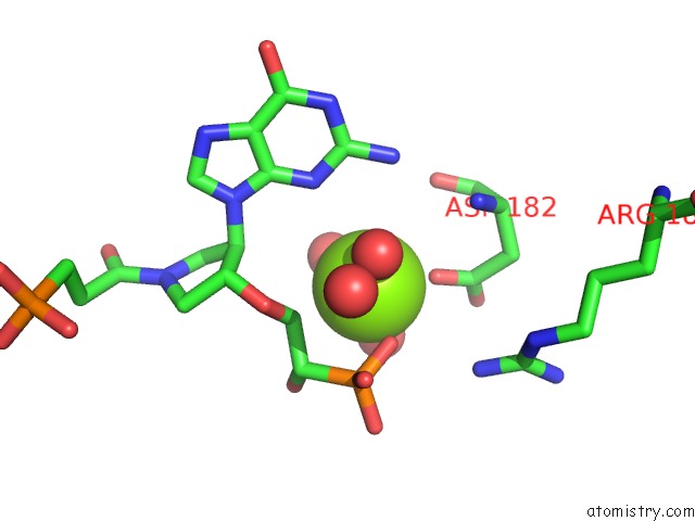
Mono view

Stereo pair view
Mono view
Stereo pair view
A full contact list of Magnesium with other atoms in the Mg binding
site number 2 of Crystal Structure of Mycobacterium Tuberculosis Hypoxanthine Guanine Phosphoribosyltransferase in Complex with (3-((3R,4R)-3-(Guanin-9- Yl)-4-((S)-2-Hydroxy-2-Phosphonoethoxy)Pyrrolidin-1-Yl)-3-Oxopropyl) Phosphonic Acid within 5.0Å range:
|
Magnesium binding site 3 out of 4 in 5kny
Go back to
Magnesium binding site 3 out
of 4 in the Crystal Structure of Mycobacterium Tuberculosis Hypoxanthine Guanine Phosphoribosyltransferase in Complex with (3-((3R,4R)-3-(Guanin-9- Yl)-4-((S)-2-Hydroxy-2-Phosphonoethoxy)Pyrrolidin-1-Yl)-3-Oxopropyl) Phosphonic Acid
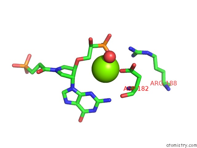
Mono view
Stereo pair view
Mono view
Stereo pair view
A full contact list of Magnesium with other atoms in the Mg binding
site number 3 of Crystal Structure of Mycobacterium Tuberculosis Hypoxanthine Guanine Phosphoribosyltransferase in Complex with (3-((3R,4R)-3-(Guanin-9- Yl)-4-((S)-2-Hydroxy-2-Phosphonoethoxy)Pyrrolidin-1-Yl)-3-Oxopropyl) Phosphonic Acid within 5.0Å range:
|
Magnesium binding site 4 out of 4 in 5kny
Go back to
Magnesium binding site 4 out
of 4 in the Crystal Structure of Mycobacterium Tuberculosis Hypoxanthine Guanine Phosphoribosyltransferase in Complex with (3-((3R,4R)-3-(Guanin-9- Yl)-4-((S)-2-Hydroxy-2-Phosphonoethoxy)Pyrrolidin-1-Yl)-3-Oxopropyl) Phosphonic Acid
Mono view
Stereo pair view
Mono view
Stereo pair view
A full contact list of Magnesium with other atoms in the Mg binding
site number 4 of Crystal Structure of Mycobacterium Tuberculosis Hypoxanthine Guanine Phosphoribosyltransferase in Complex with (3-((3R,4R)-3-(Guanin-9- Yl)-4-((S)-2-Hydroxy-2-Phosphonoethoxy)Pyrrolidin-1-Yl)-3-Oxopropyl) Phosphonic Acid within 5.0Å range:
|
Reference:
W.S.Eng,
D.Rejman,
D.T.Keough,
L.W.Guddat.
Crystal Structure of Mycobacterium Tuberculosis Hypoxanthine Guanine Phosphoribosyltransferase in Complex with Pyrrolidine Nucleoside Phosphonate To Be Published.
Page generated: Sun Sep 29 19:10:12 2024
Last articles
Fe in 2YXOFe in 2YRS
Fe in 2YXC
Fe in 2YNM
Fe in 2YVJ
Fe in 2YP1
Fe in 2YU2
Fe in 2YU1
Fe in 2YQB
Fe in 2YOO