Magnesium »
PDB 5kfv-5ktj »
5kpy »
Magnesium in PDB 5kpy: Structure of A 5-Hydroxytryptophan Aptamer
Protein crystallography data
The structure of Structure of A 5-Hydroxytryptophan Aptamer, PDB code: 5kpy
was solved by
R.T.Batey,
E.Porter,
M.Merck,
with X-Ray Crystallography technique. A brief refinement statistics is given in the table below:
| Resolution Low / High (Å) | 18.33 / 2.00 |
| Space group | C 1 2 1 |
| Cell size a, b, c (Å), α, β, γ (°) | 127.553, 26.594, 63.367, 90.00, 106.32, 90.00 |
| R / Rfree (%) | 21.7 / 25.8 |
Other elements in 5kpy:
The structure of Structure of A 5-Hydroxytryptophan Aptamer also contains other interesting chemical elements:
| Iridium | (Ir) | 12 atoms |
Magnesium Binding Sites:
The binding sites of Magnesium atom in the Structure of A 5-Hydroxytryptophan Aptamer
(pdb code 5kpy). This binding sites where shown within
5.0 Angstroms radius around Magnesium atom.
In total 6 binding sites of Magnesium where determined in the Structure of A 5-Hydroxytryptophan Aptamer, PDB code: 5kpy:
Jump to Magnesium binding site number: 1; 2; 3; 4; 5; 6;
In total 6 binding sites of Magnesium where determined in the Structure of A 5-Hydroxytryptophan Aptamer, PDB code: 5kpy:
Jump to Magnesium binding site number: 1; 2; 3; 4; 5; 6;
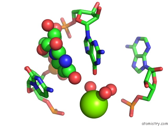
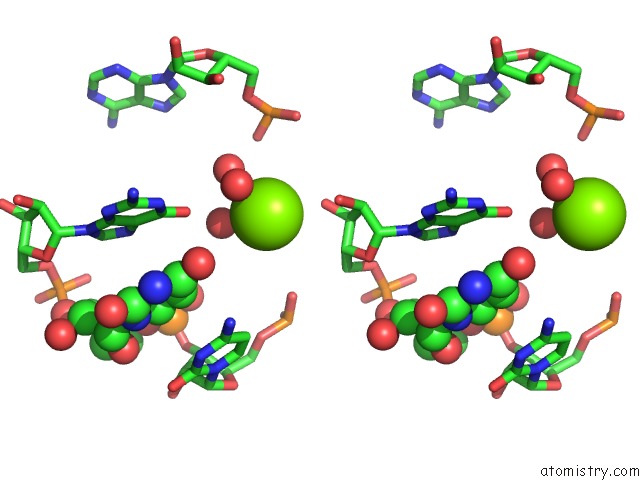
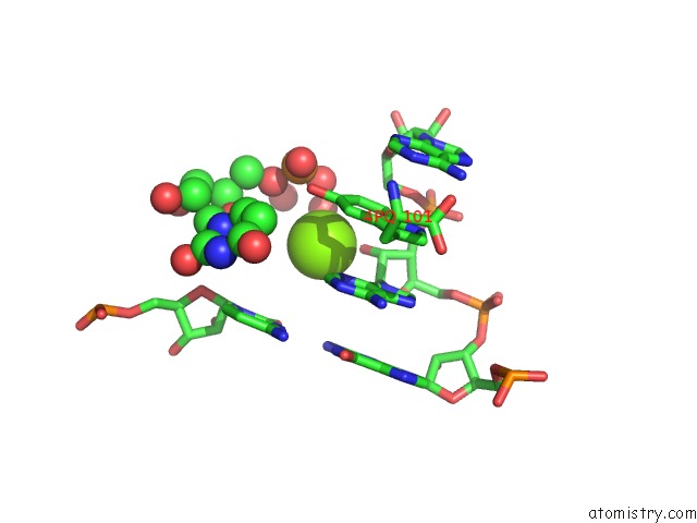
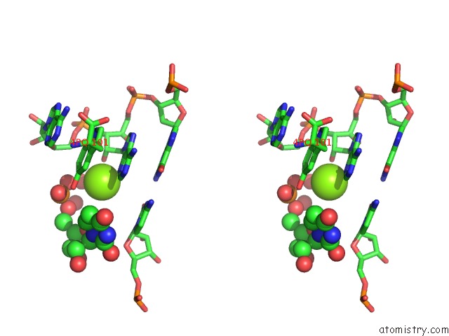
Magnesium binding site 1 out of 6 in 5kpy
Go back to
Magnesium binding site 1 out
of 6 in the Structure of A 5-Hydroxytryptophan Aptamer
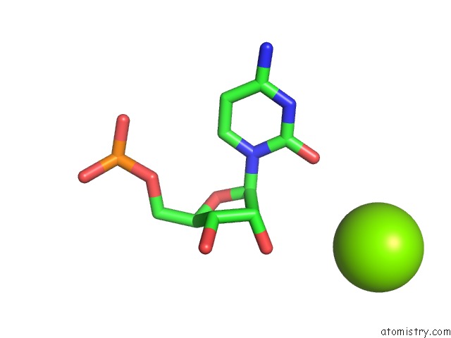
Mono view
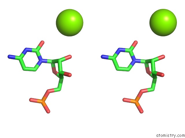
Stereo pair view
Mono view
Stereo pair view
A full contact list of Magnesium with other atoms in the Mg binding
site number 1 of Structure of A 5-Hydroxytryptophan Aptamer within 5.0Å range:
|
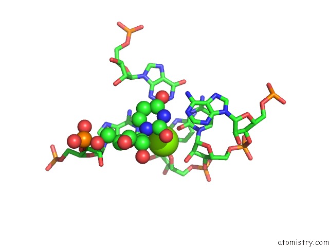
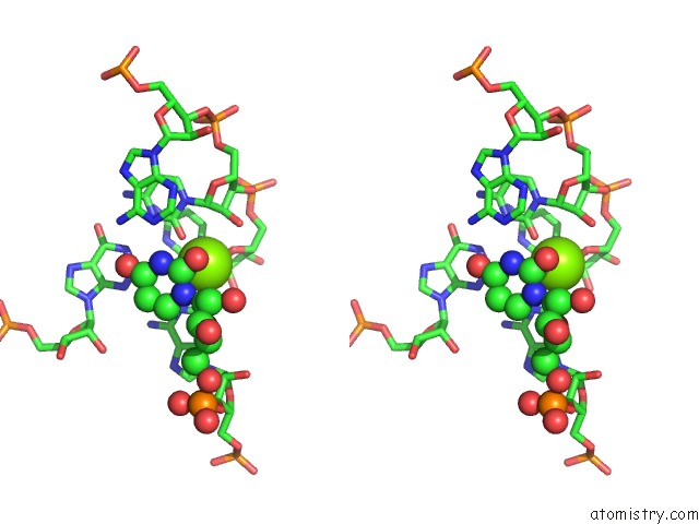
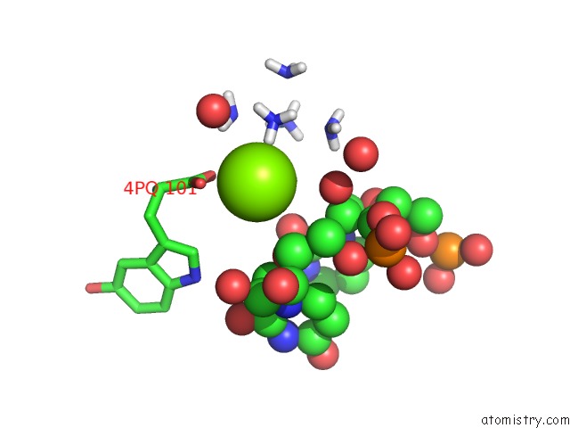
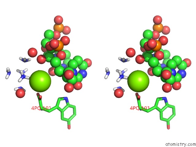
Magnesium binding site 2 out of 6 in 5kpy
Go back to
Magnesium binding site 2 out
of 6 in the Structure of A 5-Hydroxytryptophan Aptamer
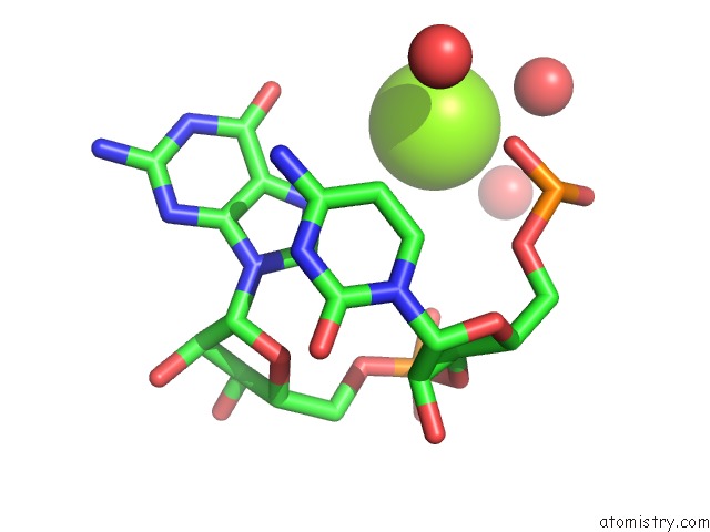
Mono view
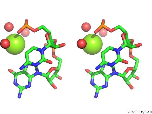
Stereo pair view
Mono view
Stereo pair view
A full contact list of Magnesium with other atoms in the Mg binding
site number 2 of Structure of A 5-Hydroxytryptophan Aptamer within 5.0Å range:
|
Magnesium binding site 3 out of 6 in 5kpy
Go back to
Magnesium binding site 3 out
of 6 in the Structure of A 5-Hydroxytryptophan Aptamer
Mono view
Stereo pair view
Mono view
Stereo pair view
A full contact list of Magnesium with other atoms in the Mg binding
site number 3 of Structure of A 5-Hydroxytryptophan Aptamer within 5.0Å range:
|
Magnesium binding site 4 out of 6 in 5kpy
Go back to
Magnesium binding site 4 out
of 6 in the Structure of A 5-Hydroxytryptophan Aptamer
Mono view
Stereo pair view
Mono view
Stereo pair view
A full contact list of Magnesium with other atoms in the Mg binding
site number 4 of Structure of A 5-Hydroxytryptophan Aptamer within 5.0Å range:
|
Magnesium binding site 5 out of 6 in 5kpy
Go back to
Magnesium binding site 5 out
of 6 in the Structure of A 5-Hydroxytryptophan Aptamer
Mono view
Stereo pair view
Mono view
Stereo pair view
A full contact list of Magnesium with other atoms in the Mg binding
site number 5 of Structure of A 5-Hydroxytryptophan Aptamer within 5.0Å range:
|
Magnesium binding site 6 out of 6 in 5kpy
Go back to
Magnesium binding site 6 out
of 6 in the Structure of A 5-Hydroxytryptophan Aptamer
Mono view
Stereo pair view
Mono view
Stereo pair view
A full contact list of Magnesium with other atoms in the Mg binding
site number 6 of Structure of A 5-Hydroxytryptophan Aptamer within 5.0Å range:
|
Reference:
E.B.Porter,
J.T.Polaski,
M.M.Morck,
R.T.Batey.
Recurrent Rna Motifs As Scaffolds For Genetically Encodable Small-Molecule Biosensors. Nat. Chem. Biol. V. 13 295 2017.
ISSN: ESSN 1552-4469
PubMed: 28092358
DOI: 10.1038/NCHEMBIO.2278
Page generated: Sun Sep 29 19:11:17 2024
ISSN: ESSN 1552-4469
PubMed: 28092358
DOI: 10.1038/NCHEMBIO.2278
Last articles
Zn in 9MJ5Zn in 9HNW
Zn in 9G0L
Zn in 9FNE
Zn in 9DZN
Zn in 9E0I
Zn in 9D32
Zn in 9DAK
Zn in 8ZXC
Zn in 8ZUF