Magnesium »
PDB 5kty-5l5u »
5l43 »
Magnesium in PDB 5l43: Structure of K26-Dcp
Protein crystallography data
The structure of Structure of K26-Dcp, PDB code: 5l43
was solved by
G.Masuyer,
K.R.Acharya,
G.J.Kramer,
B.O.Bachmann,
with X-Ray Crystallography technique. A brief refinement statistics is given in the table below:
| Resolution Low / High (Å) | 58.81 / 1.80 |
| Space group | P 1 |
| Cell size a, b, c (Å), α, β, γ (°) | 61.470, 66.936, 101.786, 97.34, 90.86, 117.31 |
| R / Rfree (%) | 17.7 / 20 |
Other elements in 5l43:
The structure of Structure of K26-Dcp also contains other interesting chemical elements:
| Zinc | (Zn) | 2 atoms |
Magnesium Binding Sites:
The binding sites of Magnesium atom in the Structure of K26-Dcp
(pdb code 5l43). This binding sites where shown within
5.0 Angstroms radius around Magnesium atom.
In total 2 binding sites of Magnesium where determined in the Structure of K26-Dcp, PDB code: 5l43:
Jump to Magnesium binding site number: 1; 2;
In total 2 binding sites of Magnesium where determined in the Structure of K26-Dcp, PDB code: 5l43:
Jump to Magnesium binding site number: 1; 2;
Magnesium binding site 1 out of 2 in 5l43
Go back to
Magnesium binding site 1 out
of 2 in the Structure of K26-Dcp
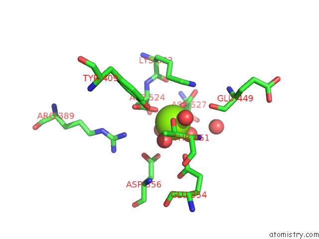
Mono view
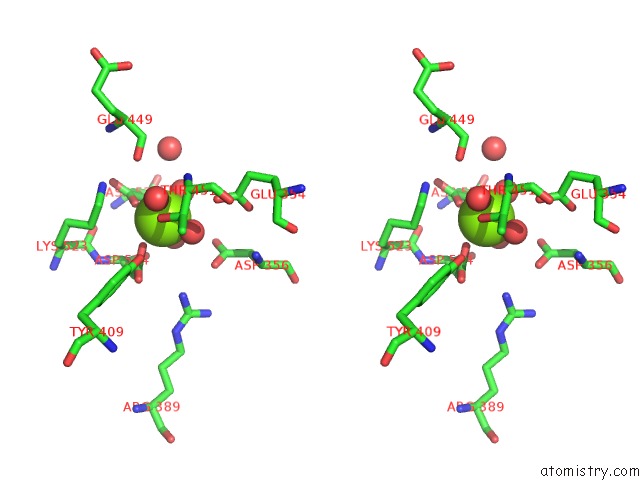
Stereo pair view
Mono view
Stereo pair view
A full contact list of Magnesium with other atoms in the Mg binding
site number 1 of Structure of K26-Dcp within 5.0Å range:
|
Magnesium binding site 2 out of 2 in 5l43
Go back to
Magnesium binding site 2 out
of 2 in the Structure of K26-Dcp
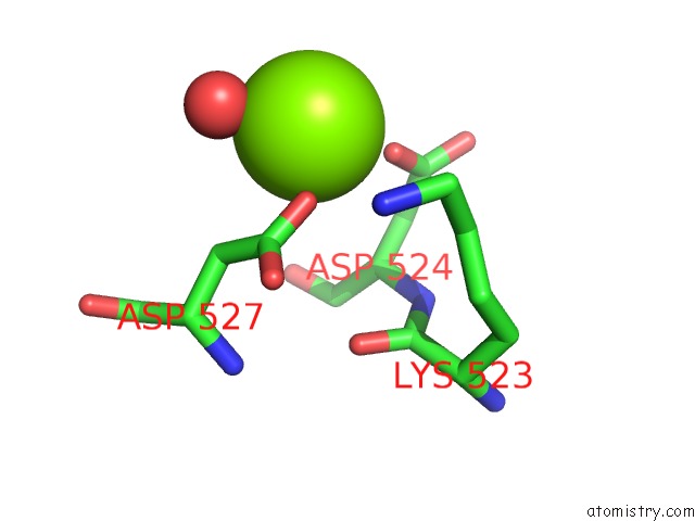
Mono view
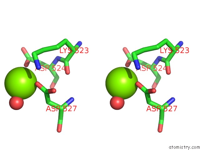
Stereo pair view
Mono view
Stereo pair view
A full contact list of Magnesium with other atoms in the Mg binding
site number 2 of Structure of K26-Dcp within 5.0Å range:
|
Reference:
G.Masuyer,
G.E.Cozier,
G.J.Kramer,
B.O.Bachmann,
K.R.Acharya.
Crystal Structure of A Peptidyl-Dipeptidase K-26-Dcp From Actinomycete in Complex with Its Natural Inhibitor. Febs J. V. 283 4357 2016.
ISSN: ISSN 1742-4658
PubMed: 27754586
DOI: 10.1111/FEBS.13928
Page generated: Sun Sep 29 19:19:01 2024
ISSN: ISSN 1742-4658
PubMed: 27754586
DOI: 10.1111/FEBS.13928
Last articles
Fe in 2YXOFe in 2YRS
Fe in 2YXC
Fe in 2YNM
Fe in 2YVJ
Fe in 2YP1
Fe in 2YU2
Fe in 2YU1
Fe in 2YQB
Fe in 2YOO