Magnesium »
PDB 5ld1-5lmn »
5lit »
Magnesium in PDB 5lit: Structure of the Dna Duplex D(Aaattt)2 with the Potential Antiparasitic Drug 6XV at 1.25 A Resolution
Protein crystallography data
The structure of Structure of the Dna Duplex D(Aaattt)2 with the Potential Antiparasitic Drug 6XV at 1.25 A Resolution, PDB code: 5lit
was solved by
C.R.Millan,
C.Dardonville,
H.P.De Koning,
N.Saperas,
J.Lourdes Campos,
with X-Ray Crystallography technique. A brief refinement statistics is given in the table below:
| Resolution Low / High (Å) | 36.14 / 1.25 |
| Space group | I 1 2 1 |
| Cell size a, b, c (Å), α, β, γ (°) | 22.330, 40.250, 72.423, 90.00, 93.67, 90.00 |
| R / Rfree (%) | 12.1 / 16.7 |
Magnesium Binding Sites:
The binding sites of Magnesium atom in the Structure of the Dna Duplex D(Aaattt)2 with the Potential Antiparasitic Drug 6XV at 1.25 A Resolution
(pdb code 5lit). This binding sites where shown within
5.0 Angstroms radius around Magnesium atom.
In total 4 binding sites of Magnesium where determined in the Structure of the Dna Duplex D(Aaattt)2 with the Potential Antiparasitic Drug 6XV at 1.25 A Resolution, PDB code: 5lit:
Jump to Magnesium binding site number: 1; 2; 3; 4;
In total 4 binding sites of Magnesium where determined in the Structure of the Dna Duplex D(Aaattt)2 with the Potential Antiparasitic Drug 6XV at 1.25 A Resolution, PDB code: 5lit:
Jump to Magnesium binding site number: 1; 2; 3; 4;
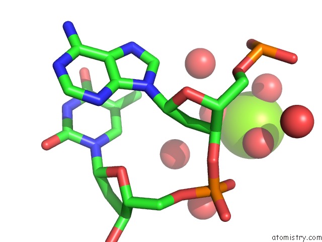
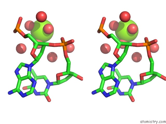
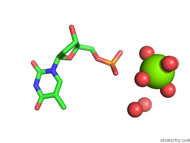
Magnesium binding site 1 out of 4 in 5lit
Go back to
Magnesium binding site 1 out
of 4 in the Structure of the Dna Duplex D(Aaattt)2 with the Potential Antiparasitic Drug 6XV at 1.25 A Resolution

Mono view

Stereo pair view
Mono view
Stereo pair view
A full contact list of Magnesium with other atoms in the Mg binding
site number 1 of Structure of the Dna Duplex D(Aaattt)2 with the Potential Antiparasitic Drug 6XV at 1.25 A Resolution within 5.0Å range:
|
Magnesium binding site 2 out of 4 in 5lit
Go back to
Magnesium binding site 2 out
of 4 in the Structure of the Dna Duplex D(Aaattt)2 with the Potential Antiparasitic Drug 6XV at 1.25 A Resolution
Mono view
Stereo pair view
Mono view
Stereo pair view
A full contact list of Magnesium with other atoms in the Mg binding
site number 2 of Structure of the Dna Duplex D(Aaattt)2 with the Potential Antiparasitic Drug 6XV at 1.25 A Resolution within 5.0Å range:
|
Magnesium binding site 3 out of 4 in 5lit
Go back to
Magnesium binding site 3 out
of 4 in the Structure of the Dna Duplex D(Aaattt)2 with the Potential Antiparasitic Drug 6XV at 1.25 A Resolution

Mono view

Stereo pair view
Mono view
Stereo pair view
A full contact list of Magnesium with other atoms in the Mg binding
site number 3 of Structure of the Dna Duplex D(Aaattt)2 with the Potential Antiparasitic Drug 6XV at 1.25 A Resolution within 5.0Å range:
|
Magnesium binding site 4 out of 4 in 5lit
Go back to
Magnesium binding site 4 out
of 4 in the Structure of the Dna Duplex D(Aaattt)2 with the Potential Antiparasitic Drug 6XV at 1.25 A Resolution
Mono view

Stereo pair view
Mono view
Stereo pair view
A full contact list of Magnesium with other atoms in the Mg binding
site number 4 of Structure of the Dna Duplex D(Aaattt)2 with the Potential Antiparasitic Drug 6XV at 1.25 A Resolution within 5.0Å range:
|
Reference:
C.R.Millan,
F.J.Acosta-Reyes,
L.Lagartera,
G.U.Ebiloma,
L.Lemgruber,
J.J.Nue Martinez,
N.Saperas,
C.Dardonville,
H.P.De Koning,
J.L.Campos.
Functional and Structural Analysis of at-Specific Minor Groove Binders That Disrupt Dna-Protein Interactions and Cause Disintegration of the Trypanosoma Brucei Kinetoplast. Nucleic Acids Res. V. 45 8378 2017.
ISSN: ESSN 1362-4962
PubMed: 28637278
DOI: 10.1093/NAR/GKX521
Page generated: Sun Sep 29 20:12:44 2024
ISSN: ESSN 1362-4962
PubMed: 28637278
DOI: 10.1093/NAR/GKX521
Last articles
Cl in 5W15Cl in 5W4M
Cl in 5W4I
Cl in 5W4L
Cl in 5W44
Cl in 5W4D
Cl in 5W2O
Cl in 5W2J
Cl in 5VZH
Cl in 5VZB