Magnesium in PDB 5m6g: Crystal Structure Glucan 1,4-Beta-Glucosidase From Saccharopolyspora Erythraea
Enzymatic activity of Crystal Structure Glucan 1,4-Beta-Glucosidase From Saccharopolyspora Erythraea
All present enzymatic activity of Crystal Structure Glucan 1,4-Beta-Glucosidase From Saccharopolyspora Erythraea:
3.2.1.74;
Protein crystallography data
The structure of Crystal Structure Glucan 1,4-Beta-Glucosidase From Saccharopolyspora Erythraea, PDB code: 5m6g
was solved by
A.Gabdulkhakov,
S.Tishchenko,
A.Lisov,
A.Leontievsky,
with X-Ray Crystallography technique. A brief refinement statistics is given in the table below:
Resolution Low / High (Å)
|
45.51 /
1.83
|
Space group
|
P 21 21 21
|
Cell size a, b, c (Å), α, β, γ (°)
|
64.292,
75.717,
113.893,
90.00,
90.00,
90.00
|
R / Rfree (%)
|
17.2 /
20.4
|
Magnesium Binding Sites:
The binding sites of Magnesium atom in the Crystal Structure Glucan 1,4-Beta-Glucosidase From Saccharopolyspora Erythraea
(pdb code 5m6g). This binding sites where shown within
5.0 Angstroms radius around Magnesium atom.
In total 6 binding sites of Magnesium where determined in the
Crystal Structure Glucan 1,4-Beta-Glucosidase From Saccharopolyspora Erythraea, PDB code: 5m6g:
Jump to Magnesium binding site number:
1;
2;
3;
4;
5;
6;
Magnesium binding site 1 out
of 6 in 5m6g
Go back to
Magnesium Binding Sites List in 5m6g
Magnesium binding site 1 out
of 6 in the Crystal Structure Glucan 1,4-Beta-Glucosidase From Saccharopolyspora Erythraea
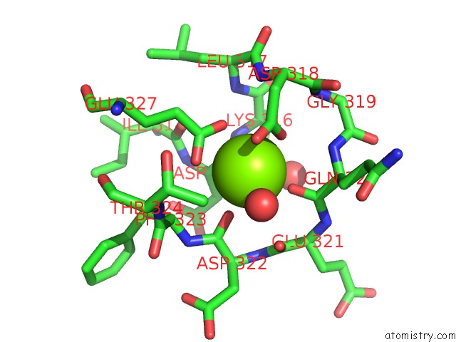
Mono view
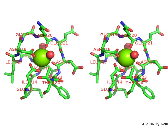
Stereo pair view
|
A full contact list of Magnesium with other atoms in the Mg binding
site number 1 of Crystal Structure Glucan 1,4-Beta-Glucosidase From Saccharopolyspora Erythraea within 5.0Å range:
probe
|
atom
|
residue
|
distance (Å)
|
B
|
Occ
|
A:Mg701
b:14.0
occ:1.00
|
O
|
A:HOH843
|
2.3
|
23.7
|
1.0
|
O
|
A:ASP315
|
2.3
|
20.8
|
1.0
|
OD1
|
A:ASP318
|
2.4
|
29.2
|
1.0
|
O
|
A:ASP322
|
2.4
|
23.9
|
1.0
|
O
|
A:GLN320
|
2.4
|
26.1
|
1.0
|
OE2
|
A:GLU327
|
2.5
|
19.5
|
1.0
|
OE1
|
A:GLU327
|
2.6
|
23.0
|
1.0
|
CD
|
A:GLU327
|
2.9
|
21.9
|
1.0
|
CG
|
A:ASP318
|
3.0
|
27.1
|
1.0
|
OD2
|
A:ASP318
|
3.1
|
26.4
|
1.0
|
C
|
A:ASP315
|
3.3
|
17.4
|
1.0
|
C
|
A:GLN320
|
3.5
|
28.2
|
1.0
|
C
|
A:ASP322
|
3.7
|
22.9
|
1.0
|
O
|
A:GLU321
|
3.7
|
27.6
|
1.0
|
CA
|
A:ASP315
|
3.8
|
18.0
|
1.0
|
N
|
A:GLN320
|
4.1
|
27.6
|
1.0
|
C
|
A:GLU321
|
4.1
|
25.1
|
1.0
|
CA
|
A:GLN320
|
4.2
|
31.0
|
1.0
|
CA
|
A:PHE323
|
4.3
|
22.6
|
1.0
|
CG
|
A:GLU327
|
4.4
|
23.4
|
1.0
|
CB
|
A:ASP318
|
4.4
|
23.5
|
1.0
|
N
|
A:ASP318
|
4.4
|
24.3
|
1.0
|
N
|
A:PHE323
|
4.4
|
26.9
|
1.0
|
N
|
A:LYS316
|
4.5
|
18.6
|
1.0
|
CB
|
A:GLN320
|
4.5
|
30.7
|
1.0
|
N
|
A:ASP322
|
4.5
|
23.5
|
1.0
|
N
|
A:GLU321
|
4.5
|
23.4
|
1.0
|
O
|
A:ILE314
|
4.5
|
17.9
|
1.0
|
CB
|
A:ASP315
|
4.5
|
17.5
|
1.0
|
O
|
A:HOH1028
|
4.5
|
26.3
|
1.0
|
CA
|
A:ASP322
|
4.7
|
26.1
|
1.0
|
CA
|
A:GLU321
|
4.8
|
29.0
|
1.0
|
N
|
A:THR324
|
4.8
|
20.9
|
1.0
|
CA
|
A:ASP318
|
4.8
|
26.0
|
1.0
|
CG2
|
A:THR324
|
4.8
|
19.4
|
1.0
|
N
|
A:LEU317
|
4.9
|
21.9
|
1.0
|
CA
|
A:LYS316
|
4.9
|
17.8
|
1.0
|
N
|
A:GLY319
|
4.9
|
29.4
|
1.0
|
C
|
A:PHE323
|
4.9
|
21.3
|
1.0
|
C
|
A:ASP318
|
5.0
|
30.1
|
1.0
|
|
Magnesium binding site 2 out
of 6 in 5m6g
Go back to
Magnesium Binding Sites List in 5m6g
Magnesium binding site 2 out
of 6 in the Crystal Structure Glucan 1,4-Beta-Glucosidase From Saccharopolyspora Erythraea
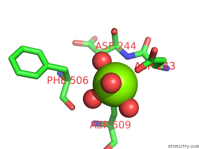
Mono view
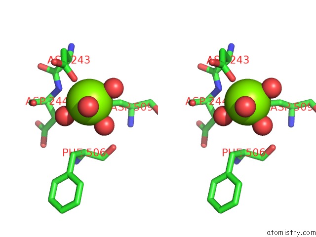
Stereo pair view
|
A full contact list of Magnesium with other atoms in the Mg binding
site number 2 of Crystal Structure Glucan 1,4-Beta-Glucosidase From Saccharopolyspora Erythraea within 5.0Å range:
probe
|
atom
|
residue
|
distance (Å)
|
B
|
Occ
|
A:Mg702
b:19.4
occ:1.00
|
O
|
A:HOH962
|
2.0
|
20.6
|
1.0
|
OD1
|
A:ASP243
|
2.1
|
19.2
|
1.0
|
O
|
A:HOH1032
|
2.1
|
16.6
|
1.0
|
O
|
A:HOH919
|
2.1
|
19.7
|
1.0
|
OD2
|
A:ASP509
|
2.1
|
14.9
|
1.0
|
O
|
A:HOH1054
|
2.1
|
22.7
|
1.0
|
CG
|
A:ASP509
|
3.2
|
15.8
|
1.0
|
CG
|
A:ASP243
|
3.3
|
22.1
|
1.0
|
CB
|
A:ASP509
|
3.5
|
14.6
|
1.0
|
N
|
A:ASP244
|
3.9
|
15.5
|
1.0
|
OD2
|
A:ASP243
|
4.0
|
24.7
|
1.0
|
CA
|
A:ASP243
|
4.3
|
17.8
|
1.0
|
OD1
|
A:ASP509
|
4.3
|
11.9
|
1.0
|
CB
|
A:ASP243
|
4.3
|
16.3
|
1.0
|
O
|
A:ASP244
|
4.4
|
16.2
|
1.0
|
O
|
A:PHE506
|
4.4
|
15.9
|
1.0
|
CB
|
A:PHE506
|
4.5
|
15.1
|
1.0
|
C
|
A:ASP243
|
4.5
|
17.0
|
1.0
|
CB
|
A:ASP244
|
4.6
|
11.9
|
1.0
|
CA
|
A:ASP244
|
4.7
|
14.6
|
1.0
|
CA
|
A:PHE506
|
4.8
|
17.4
|
1.0
|
|
Magnesium binding site 3 out
of 6 in 5m6g
Go back to
Magnesium Binding Sites List in 5m6g
Magnesium binding site 3 out
of 6 in the Crystal Structure Glucan 1,4-Beta-Glucosidase From Saccharopolyspora Erythraea
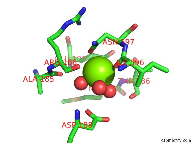
Mono view
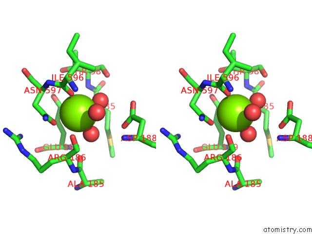
Stereo pair view
|
A full contact list of Magnesium with other atoms in the Mg binding
site number 3 of Crystal Structure Glucan 1,4-Beta-Glucosidase From Saccharopolyspora Erythraea within 5.0Å range:
probe
|
atom
|
residue
|
distance (Å)
|
B
|
Occ
|
A:Mg703
b:21.5
occ:1.00
|
OE1
|
A:GLU199
|
2.2
|
12.9
|
1.0
|
O
|
A:HOH831
|
2.2
|
19.0
|
1.0
|
O
|
A:HOH916
|
2.3
|
12.7
|
1.0
|
O
|
A:ILE596
|
2.3
|
14.5
|
1.0
|
OD1
|
A:ASN597
|
2.4
|
15.0
|
1.0
|
CG
|
A:ASN597
|
3.2
|
16.2
|
1.0
|
CD
|
A:GLU199
|
3.2
|
17.9
|
1.0
|
O
|
A:HOH1020
|
3.4
|
12.8
|
1.0
|
C
|
A:ILE596
|
3.5
|
14.7
|
1.0
|
CB
|
A:GLU199
|
3.5
|
16.3
|
1.0
|
CA
|
A:ASN597
|
3.9
|
15.0
|
1.0
|
ND2
|
A:ASN597
|
3.9
|
14.0
|
1.0
|
O
|
A:ARG186
|
3.9
|
17.8
|
1.0
|
CG
|
A:GLU199
|
4.0
|
13.5
|
1.0
|
CB
|
A:ASN597
|
4.1
|
16.0
|
1.0
|
OE2
|
A:GLU199
|
4.1
|
15.8
|
1.0
|
N
|
A:ASN597
|
4.2
|
13.9
|
1.0
|
O
|
A:ALA185
|
4.2
|
14.2
|
1.0
|
N
|
A:THR586
|
4.2
|
15.3
|
1.0
|
C
|
A:ARG186
|
4.3
|
15.1
|
1.0
|
OD1
|
A:ASP188
|
4.3
|
24.1
|
1.0
|
CA
|
A:ARG186
|
4.4
|
12.6
|
1.0
|
CG
|
A:MET585
|
4.4
|
19.2
|
1.0
|
CB
|
A:MET585
|
4.5
|
15.6
|
1.0
|
O
|
A:THR586
|
4.6
|
14.8
|
1.0
|
N
|
A:ILE596
|
4.6
|
14.8
|
1.0
|
CB
|
A:THR586
|
4.6
|
14.1
|
1.0
|
CA
|
A:ILE596
|
4.6
|
13.8
|
1.0
|
CG2
|
A:ILE596
|
4.8
|
12.3
|
1.0
|
CA
|
A:MET585
|
4.8
|
13.7
|
1.0
|
CA
|
A:THR586
|
4.9
|
14.9
|
1.0
|
CA
|
A:GLU199
|
4.9
|
17.9
|
1.0
|
CG
|
A:ASP188
|
5.0
|
19.9
|
1.0
|
C
|
A:MET585
|
5.0
|
17.0
|
1.0
|
|
Magnesium binding site 4 out
of 6 in 5m6g
Go back to
Magnesium Binding Sites List in 5m6g
Magnesium binding site 4 out
of 6 in the Crystal Structure Glucan 1,4-Beta-Glucosidase From Saccharopolyspora Erythraea
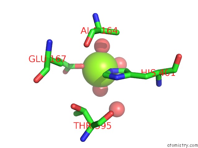
Mono view
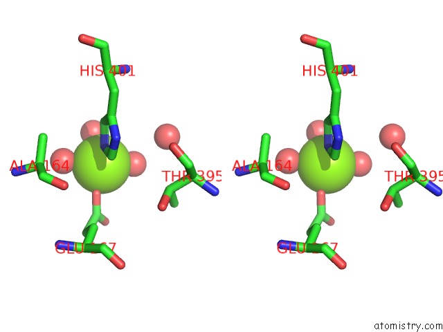
Stereo pair view
|
A full contact list of Magnesium with other atoms in the Mg binding
site number 4 of Crystal Structure Glucan 1,4-Beta-Glucosidase From Saccharopolyspora Erythraea within 5.0Å range:
probe
|
atom
|
residue
|
distance (Å)
|
B
|
Occ
|
A:Mg704
b:27.5
occ:1.00
|
O
|
A:HOH1051
|
1.9
|
25.6
|
1.0
|
OE1
|
A:GLU167
|
1.9
|
28.0
|
1.0
|
O
|
A:HOH1040
|
2.1
|
30.6
|
1.0
|
NE2
|
A:HIS401
|
2.2
|
18.1
|
1.0
|
O
|
A:HOH937
|
2.2
|
33.3
|
1.0
|
O
|
A:HOH1052
|
2.3
|
30.5
|
1.0
|
CD
|
A:GLU167
|
3.0
|
31.1
|
1.0
|
CE1
|
A:HIS401
|
3.1
|
26.2
|
1.0
|
CD2
|
A:HIS401
|
3.2
|
15.6
|
1.0
|
OE2
|
A:GLU167
|
3.4
|
34.5
|
1.0
|
O
|
A:HOH860
|
4.1
|
25.4
|
1.0
|
ND1
|
A:HIS401
|
4.3
|
18.6
|
1.0
|
CG
|
A:GLU167
|
4.3
|
23.8
|
1.0
|
CG
|
A:HIS401
|
4.3
|
18.1
|
1.0
|
CB
|
A:GLU167
|
4.7
|
22.9
|
1.0
|
CA
|
A:ALA164
|
4.8
|
13.9
|
1.0
|
CB
|
A:THR395
|
4.9
|
20.3
|
1.0
|
CB
|
A:ALA164
|
4.9
|
15.2
|
1.0
|
CG2
|
A:THR395
|
4.9
|
20.0
|
1.0
|
|
Magnesium binding site 5 out
of 6 in 5m6g
Go back to
Magnesium Binding Sites List in 5m6g
Magnesium binding site 5 out
of 6 in the Crystal Structure Glucan 1,4-Beta-Glucosidase From Saccharopolyspora Erythraea
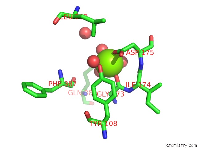
Mono view
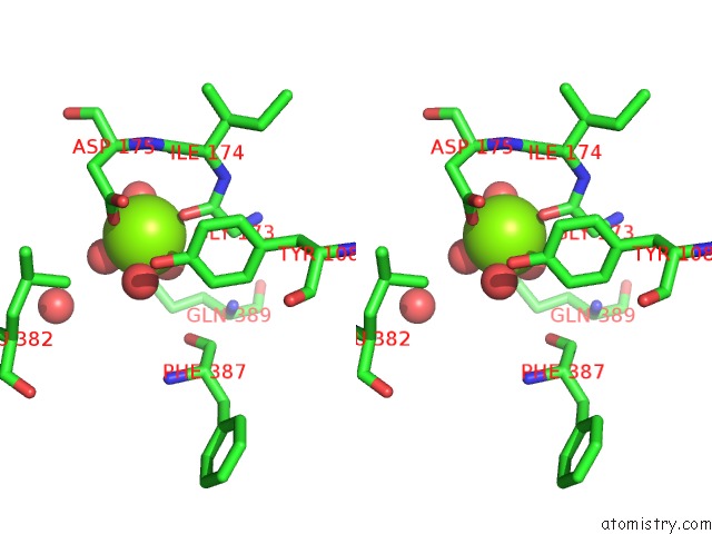
Stereo pair view
|
A full contact list of Magnesium with other atoms in the Mg binding
site number 5 of Crystal Structure Glucan 1,4-Beta-Glucosidase From Saccharopolyspora Erythraea within 5.0Å range:
probe
|
atom
|
residue
|
distance (Å)
|
B
|
Occ
|
A:Mg705
b:21.2
occ:1.00
|
O
|
A:HOH999
|
2.0
|
20.5
|
1.0
|
O
|
A:HOH1021
|
2.0
|
24.8
|
1.0
|
O
|
A:GLY173
|
2.0
|
13.7
|
1.0
|
O
|
A:HOH921
|
2.1
|
15.2
|
1.0
|
OD1
|
A:ASP175
|
2.2
|
16.2
|
1.0
|
O
|
A:HOH866
|
2.2
|
20.7
|
1.0
|
C
|
A:GLY173
|
3.1
|
15.6
|
1.0
|
CG
|
A:ASP175
|
3.3
|
17.3
|
1.0
|
CA
|
A:GLY173
|
3.7
|
14.9
|
1.0
|
OH
|
A:TYR108
|
3.8
|
13.7
|
1.0
|
O
|
A:ILE174
|
3.8
|
13.7
|
1.0
|
C
|
A:ILE174
|
3.9
|
13.6
|
1.0
|
OD2
|
A:ASP175
|
4.0
|
14.0
|
1.0
|
CA
|
A:ASP175
|
4.0
|
10.3
|
1.0
|
CB
|
A:ASP175
|
4.1
|
14.9
|
1.0
|
N
|
A:ASP175
|
4.1
|
12.1
|
1.0
|
N
|
A:ILE174
|
4.2
|
13.2
|
1.0
|
CZ
|
A:TYR108
|
4.2
|
16.5
|
1.0
|
OE1
|
A:GLN389
|
4.4
|
34.4
|
1.0
|
CA
|
A:ILE174
|
4.5
|
13.8
|
1.0
|
CD2
|
A:LEU382
|
4.5
|
19.0
|
1.0
|
CE2
|
A:TYR108
|
4.6
|
15.4
|
1.0
|
CE1
|
A:TYR108
|
4.7
|
14.8
|
1.0
|
O
|
A:HOH918
|
4.8
|
22.8
|
1.0
|
O
|
A:PHE387
|
4.8
|
18.4
|
1.0
|
N
|
A:GLY173
|
4.9
|
16.5
|
1.0
|
|
Magnesium binding site 6 out
of 6 in 5m6g
Go back to
Magnesium Binding Sites List in 5m6g
Magnesium binding site 6 out
of 6 in the Crystal Structure Glucan 1,4-Beta-Glucosidase From Saccharopolyspora Erythraea
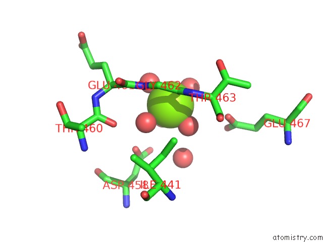
Mono view
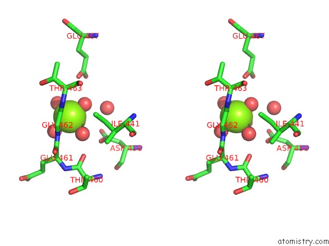
Stereo pair view
|
A full contact list of Magnesium with other atoms in the Mg binding
site number 6 of Crystal Structure Glucan 1,4-Beta-Glucosidase From Saccharopolyspora Erythraea within 5.0Å range:
probe
|
atom
|
residue
|
distance (Å)
|
B
|
Occ
|
A:Mg706
b:27.9
occ:1.00
|
O
|
A:HOH1025
|
2.0
|
16.4
|
1.0
|
O
|
A:HOH964
|
2.1
|
28.8
|
1.0
|
O
|
A:HOH883
|
2.1
|
22.5
|
1.0
|
O
|
A:GLY462
|
2.2
|
14.3
|
1.0
|
O
|
A:HOH1030
|
2.2
|
19.4
|
1.0
|
O
|
A:HOH805
|
2.3
|
21.2
|
1.0
|
C
|
A:GLY462
|
3.3
|
13.8
|
1.0
|
N
|
A:GLY462
|
3.6
|
13.0
|
1.0
|
CA
|
A:GLY462
|
4.0
|
14.6
|
1.0
|
O
|
A:HOH993
|
4.1
|
21.2
|
1.0
|
OD1
|
A:ASP458
|
4.2
|
23.3
|
1.0
|
N
|
A:THR463
|
4.3
|
13.0
|
1.0
|
OE1
|
A:GLU467
|
4.3
|
15.8
|
1.0
|
OD2
|
A:ASP458
|
4.3
|
32.9
|
1.0
|
CD1
|
A:ILE441
|
4.5
|
13.6
|
1.0
|
CA
|
A:THR463
|
4.5
|
13.7
|
1.0
|
CG1
|
A:ILE441
|
4.6
|
17.6
|
1.0
|
C
|
A:GLU461
|
4.7
|
17.4
|
1.0
|
CG
|
A:ASP458
|
4.7
|
23.3
|
1.0
|
O
|
A:THR460
|
4.7
|
17.9
|
1.0
|
CA
|
A:GLU461
|
4.8
|
17.1
|
1.0
|
OE2
|
A:GLU467
|
4.9
|
15.9
|
1.0
|
|
Reference:
A.Gabdulkhakov,
S.Tishchenko.
Crystal Structure Glucan 1,4-Beta-Glucosidase From Saccharopolyspora Erythraea To Be Published.
Page generated: Sun Sep 29 21:13:54 2024
|