Magnesium »
PDB 5m5l-5mh5 »
5m6z »
Magnesium in PDB 5m6z: The X-Ray Structure of Human M189I Pgk-1 Mutant in Partially Closed Conformation
Enzymatic activity of The X-Ray Structure of Human M189I Pgk-1 Mutant in Partially Closed Conformation
All present enzymatic activity of The X-Ray Structure of Human M189I Pgk-1 Mutant in Partially Closed Conformation:
2.7.2.3;
2.7.2.3;
Protein crystallography data
The structure of The X-Ray Structure of Human M189I Pgk-1 Mutant in Partially Closed Conformation, PDB code: 5m6z
was solved by
A.Ilari,
A.Fiorillo,
M.Petrosino,
A.Cipollone,
with X-Ray Crystallography technique. A brief refinement statistics is given in the table below:
| Resolution Low / High (Å) | 53.06 / 1.67 |
| Space group | P 1 21 1 |
| Cell size a, b, c (Å), α, β, γ (°) | 35.880, 106.121, 50.472, 90.00, 98.13, 90.00 |
| R / Rfree (%) | 18.3 / 22.3 |
Magnesium Binding Sites:
The binding sites of Magnesium atom in the The X-Ray Structure of Human M189I Pgk-1 Mutant in Partially Closed Conformation
(pdb code 5m6z). This binding sites where shown within
5.0 Angstroms radius around Magnesium atom.
In total 2 binding sites of Magnesium where determined in the The X-Ray Structure of Human M189I Pgk-1 Mutant in Partially Closed Conformation, PDB code: 5m6z:
Jump to Magnesium binding site number: 1; 2;
In total 2 binding sites of Magnesium where determined in the The X-Ray Structure of Human M189I Pgk-1 Mutant in Partially Closed Conformation, PDB code: 5m6z:
Jump to Magnesium binding site number: 1; 2;
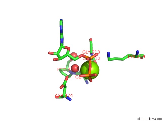
Magnesium binding site 1 out of 2 in 5m6z
Go back to
Magnesium binding site 1 out
of 2 in the The X-Ray Structure of Human M189I Pgk-1 Mutant in Partially Closed Conformation
Mono view

Stereo pair view
Mono view
Stereo pair view
A full contact list of Magnesium with other atoms in the Mg binding
site number 1 of The X-Ray Structure of Human M189I Pgk-1 Mutant in Partially Closed Conformation within 5.0Å range:
|
Magnesium binding site 2 out of 2 in 5m6z
Go back to
Magnesium binding site 2 out
of 2 in the The X-Ray Structure of Human M189I Pgk-1 Mutant in Partially Closed Conformation

Mono view

Stereo pair view
Mono view
Stereo pair view
A full contact list of Magnesium with other atoms in the Mg binding
site number 2 of The X-Ray Structure of Human M189I Pgk-1 Mutant in Partially Closed Conformation within 5.0Å range:
|
Reference:
A.Fiorillo,
M.Petrosino,
A.Ilari,
A.Pasquo,
A.Cipollone,
M.Maggi,
R.Chiaraluce,
V.Consalvi.
The Phosphoglycerate Kinase 1 Variants Found in Carcinoma Cells Display Different Catalytic Activity and Conformational Stability Compared to the Native Enzyme. Plos One V. 13 99191 2018.
ISSN: ESSN 1932-6203
PubMed: 29995887
DOI: 10.1371/JOURNAL.PONE.0199191
Page generated: Sun Sep 29 21:13:54 2024
ISSN: ESSN 1932-6203
PubMed: 29995887
DOI: 10.1371/JOURNAL.PONE.0199191
Last articles
Zn in 9MJ5Zn in 9HNW
Zn in 9G0L
Zn in 9FNE
Zn in 9DZN
Zn in 9E0I
Zn in 9D32
Zn in 9DAK
Zn in 8ZXC
Zn in 8ZUF